Automated CT Liver Volumetry by Use of Three-Dimensional Fast-Marching and Level-Set Segmentation
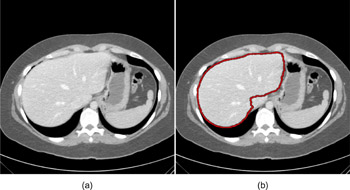
We developed an automated scheme for segmenting and calculating liver volume in hepatic CT by means of 3D fast-marching and level-set segmentation algorithms. Automatic liver segmentation on hepatic CT images is challenging because the liver often abuts other organs of similar density. Our purpose was to develop an automated liver segmentation scheme based on a 3D level-set algorithm for measuring liver volumes. Hepatic CT scans of eighteen prospective liver donors were obtained under a liver transplant protocol. Scans were acquired with a multi-detector CT system with a 16-, 40-, or 64-channel detector scanner (Brilliance, Philips Medical Systems, Netherlands). We developed an automated liver segmentation scheme for volumetry of the liver in CT. Our scheme consisted of five steps. First, a 3D anisotropic diffusion smoothing filter was applied to CT images for removing noise while preserving the structures in the liver, followed by a gradient magnitude filter for enhancing liver edges. A nonlinear gray-scale enhancement filter was applied to the gradient magnitude image for further enhancing the boundary of the liver. By use of the enhanced gradient magnitude image as a speed function, a 3D fast-marching algorithm generated an initial surface that roughly estimated the shape of the liver. A 3D level-set segmentation algorithm refined the initial surface so as to fit the liver boundary more accurately (see the figure for a segmentation result). The liver volume was calculated based on the refined liver surface. Automated volumes were compared to manually determined liver volumes. The mean liver volume obtained with our scheme was 1598 cc (range: 1002-2415 cc), whereas the mean manual volume was 1,535 cc (range: 1,007-2,435 cc). The mean absolute difference between automated and manual volumes was 128 (9.5%) 119 (9.4%) cc. The two volumetrics reached excellent agreement (the intra-class correlation coefficient was 0.89) with no statistically significant difference (P=0.13). The processing time by the automated method was 2-5 min. per case (Intel, Xeon, 2.7 GHz), whereas that by manual segmentation was approximately 50-60 min. per case. CT liver volumetrics based on an automated scheme agreed excellently with manual volumetrics and required substantially less completion time. Thus, our automated scheme provides an efficient and accurate way of measuring liver volumes in CT; thus, it would be useful for radiologists in their measurement of liver volumes.

Illustration of automated liver segmentation. (a) Original CT liver image. (b) Segmented liver obtained with our automated liver segmentation method (Red curve).

