BMAI
バイオメディカルAI研究ユニット
バイオメディカルAI研究ユニット
Ja

「NEDO モジュール型モデルによる深層学習のホワイトボックス化 成果紹介」が公開されました。 Read more...

バイオメディカルAI研究ユニットは2021年6月に設立されました。 Read more...
研究歴:
1993年- (株) 日立メディコ 技術研究所
1997年- 愛知県立大学 情報科学部
2001年- シカゴ大学 放射線学科/大学院医用物理学研究科/がん研究センター
2014年- イリノイ工科大学 医用画像研究所/電気電子工学研究科
2017年- 東京工業大学 科学技術創成研 Read more...
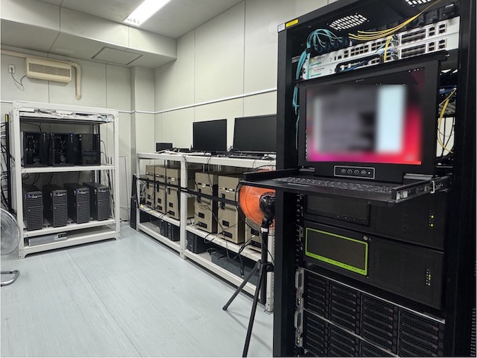
サーバー室
研究用GPUサーバー 21台
- i9-10980XE 18-Core/3.0GHz; RTX A6000x2; 128GB MEM; SSD 3.8TBx4 HDD 18TBx2
- i9-10940X 14-Core/3.3GHz; RTX 3090×2; 64GB MEM; SSD 960GB
- i9-10940X 14-Core/3.3GHz; RTX A6000; 64GB MEM; SSD 480GB
- EPYC 7502P 32-Core/2.5GHz; RTX A6000x Read more...

「JST 未来社会創造事業 成果集」が公開されました。 Read more...
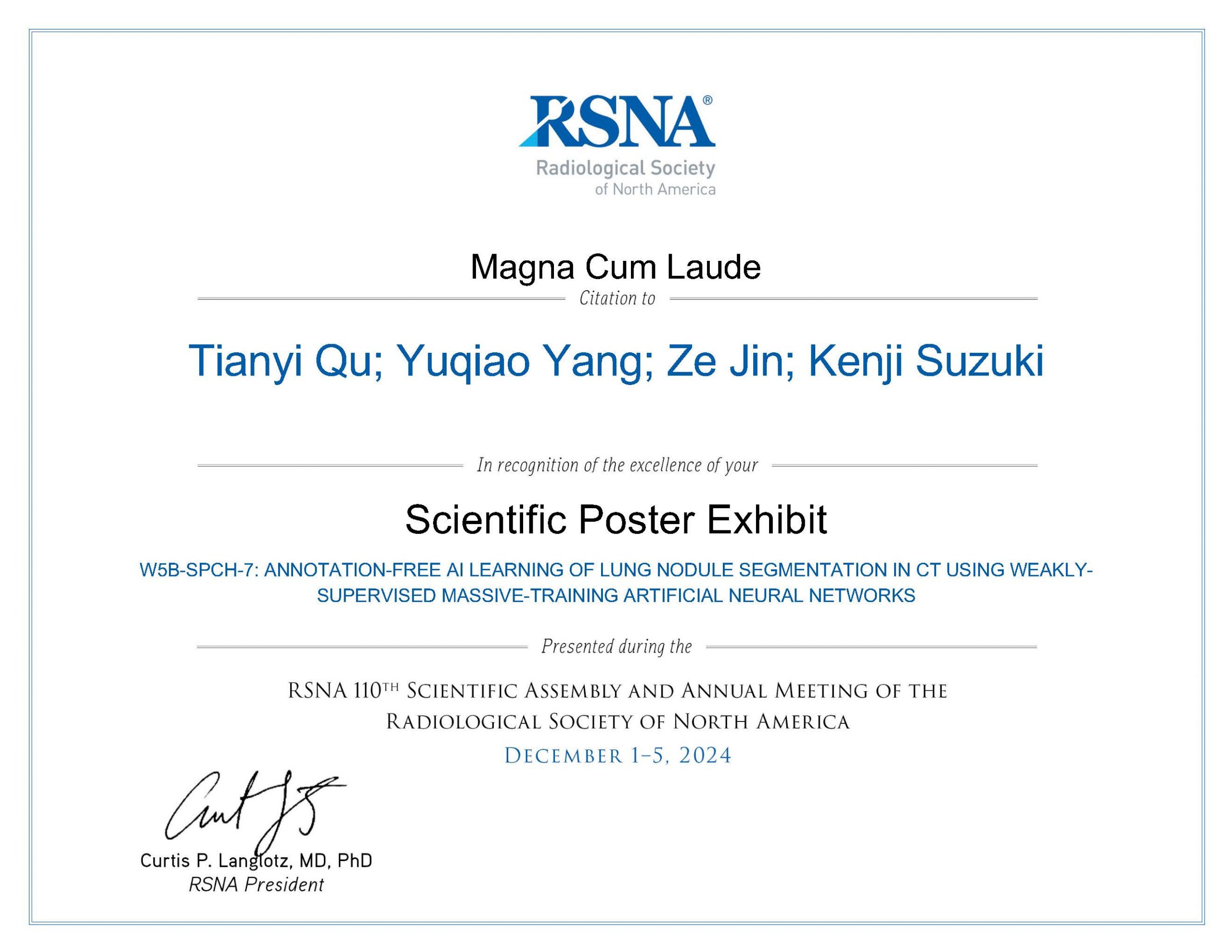
新エネルギー・産業技術総合開発機構(NEDO)による人と共に進化する次世代人工知能に関する技術開発事業の「モジュール型モデルによる深層学習のホワイトボックス化」プロジェクトに関連する発表が、RSNA 2024で最高賞の Magna Cum Laude を受賞しました! Read more...
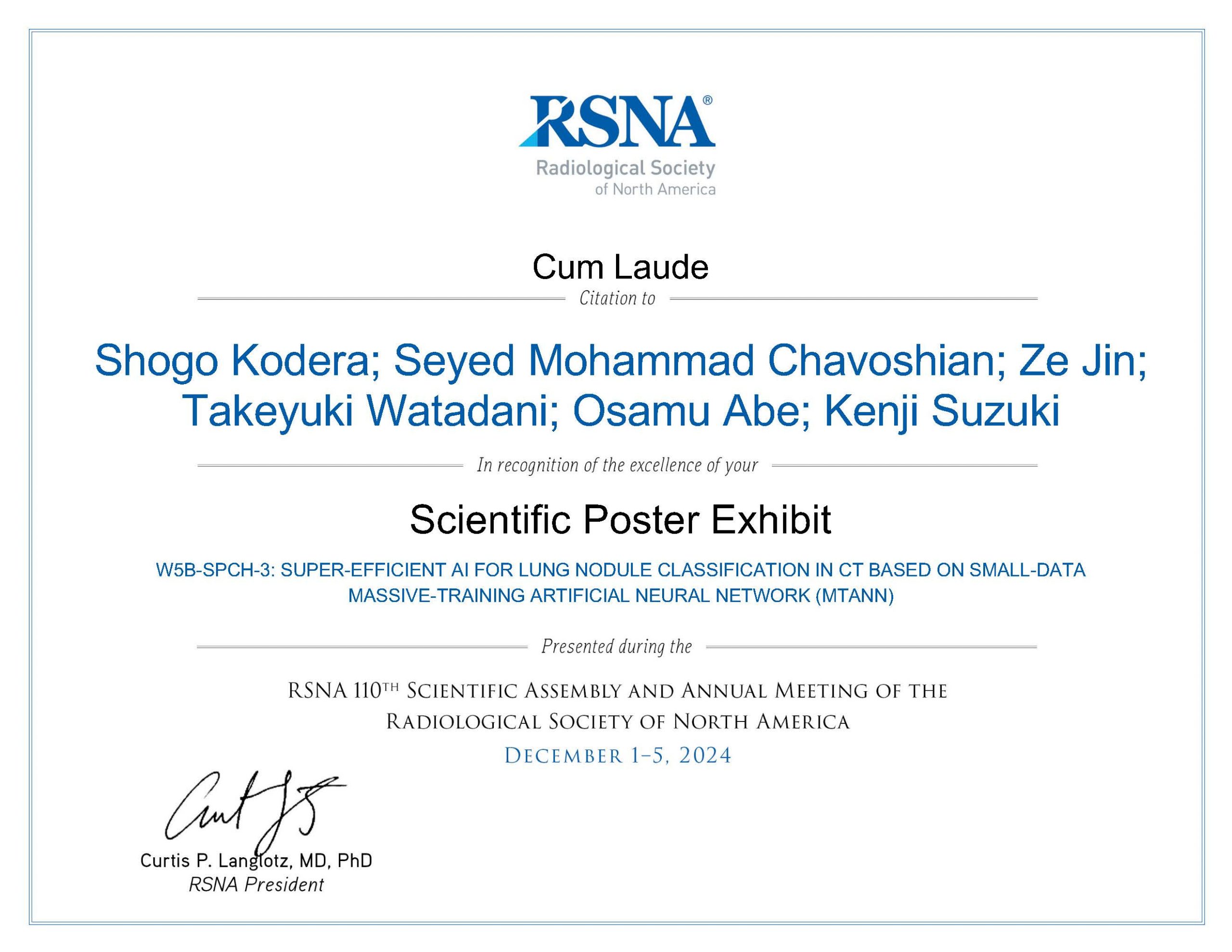
国立研究開発法人科学技術振興機構(JST)未来社会創造事業による「Engineerable AI技術による機械学習応用システムの高信頼化」プロジェクトに関連する発表が、RSNA 2024で最高に次ぐ Cum Laude を受賞しました! Read more...
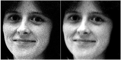
The conventional noise-reduction filters tend to blur the edge information while noise is reduced. To address this issue, we developed a supervised nonlinear filter based on an artificial neural network (ANN), called a “neural filter,” for reduction of noise in images. The neural filter is trained with input images and the corresponding teaching images. To reduce noise in images, we created Read more...
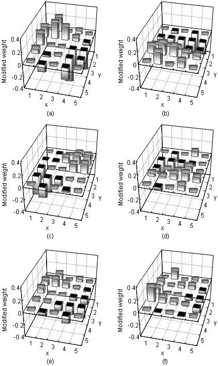
In order to gain insight into the internal presentation of a trained neural edge enhancer, we developed an analysis method for the nonlinear kernel of a trained neural edge enhancer. We trained a neural edge enhancer to enhance edges in noisy images. Our analysis method was applied to the trained neural edge enhancer with a five-by-five-pixel input kernel. Six graphs obtained by the analysis, w Read more...