
BMAI
Biomedical Artificial
Intelligence Research Unit

Biomedical Artificial
Intelligence Research Unit
En
IEEE Transactions on Pattern Analysis and Machine Intelligence
20.8
IEEE Transactions on Image Processing
10.8
Pattern Recognition
7.5
Knowledge-Based Systems
7.2
IEEE Transactions on Signal Processing
4.6
IEEE Transactions on Medical Imaging
8.9
IEEE Transactions on Biomedical Engineering
4.4
IEEE Journal of Biomedical and Health Informatics
6.7
European Journal of Nuclear Medicine and Molecular Imaging
8.6
Radiology
12.1
Liver Transplantation
4.7
Journal of Magnetic Resonance Imaging
3.3
Journal of Neural Engineering
3.7
PLoS ONE
2.9
Physics in Medicine and Biology
3.3
Medical Physics
3.2
Computer Vision and Image Understanding
4.3
American Journal of Roentgenology
4.7
Quantitative Imaging in Medicine and Surgery
2.9
| All | Since 2019 | |
|---|---|---|
| Citations | 6800 | 6376 |
| h-index | 63 | 40 |
| i10-index | 204 | 107 |
ID
Title
1
Yang Y., Sato M., Jin Z., and Suzuki K.: Patch-based Deep-learning Model with Limited Training Dataset for Liver Tumor Segmentation in Contrast-enhanced Hepatic Computed Tomography, IEEE Access 13: 86863-86873, 2025., 2025.
2
Rahmaniar W., Deng Z., Yang Y., Jin Z., and Suzuki K.: Future of the Medical World: Collaborative Medical Imaging AI with Federated Learning, IEEE Consumer Electronics Magazine, 2025.
3
Deng Z., Yang Y., and Suzuki K.: Federated Active Learning Framework for Efficient Annotation Strategy in Skin-Lesion Classification. Journal of Investigative Dermatology 145(2), 303-311, 2025.
4
Ji C., Oshima K., Urata T., Kimura F., Ishii K., Uehara T., Suzuki K., Takeyama S., and Yamaguchi M.: Transformation from Hematoxylin-and-eosin Staining to Ki-67 Immunohistochemistry Digital Staining Images Using Deep Learning: Experimental Validation on the Labeling Index. Journal of Medical Imaging 11(4), 047501, 2024.
5
Samala R. K., Drukker K., Shukla-Dave A., Chan H. P., Sahiner B., Petrick N., Greenspan H., Mahmood U., Summers R. M., Tourassi G., Deserno T. M., Regge D., Nappi J. J., Yoshida H., Huo Z., Chen Q., Vergara D., Cha K. H., Mazurchuk R., Grizzard K. T., Huisman H., Morra L., Suzuki K., Armato, III S. G., and Hadjiiski L.: AI and machine learning in medical imaging: key points from development to translation. BJR Artificial Intelligence 1(1): ubae006, 2024.
6
Mahmood U., Shukla-Dave A., Chan H. P., Drukker K., Samala R. K., Chen Q., Vergara D., Greenspan H., Petrick N., Sahiner B., Huo Z., Summers R. M., Cha K. H., Tourassi G., Deserno T. M., Grizzard K. T., Nappi J. J., Yoshida H., Regge D., Mazurchuk R., Suzuki K., Morra L., Huisman H., Armato, III S. G., and Hadjiiski L.: Artificial intelligence in medicine: mitigating risks and maximizing benefits via quality assurance, quality control, and acceptance testing. BJR Artificial Intelligence 1(1): ubae003, 2024.
7
Pavarut S., Preedanan W., Kumazawa I., Suzuki K., Kobayashi M., Tanaka H., Ishioka J., Matsuoka Y., and Fujii Y.: Improving Kidney Tumor Classification With Multi-Modal Medical Images Recovered Partially by Conditional CycleGAN. IEEE Access 11: 146250 – 146261, 2023.
8
Rahmaniar W., Suzuki K., and Lin T.L.: Auto-CA: Automated Cobb Angle Measurement Based on Vertebrae Detection for Assessment of Spinal Curvature Deformity. IEEE Transactions on Biomedical Engineering: 1-10, 2023. Top 10% paper
9
Shishido T. Ono Y., Kumazawa I. Iwai I., and Suzuki K.: Artificial intelligence model substantially improves stratum corneum moisture content prediction from visible-light skin images and skin feature factors. Skin Research and Technology 29(8): e13414, 2023.
10
Preedanan W., Suzuki K., Kondo T., Kobayashi M., Tanaka H., Ishioka J., Matsuoka Y., Fujii Y., and Kumazawa I.: Urinary Stones Segmentation in Abdominal X-Ray Images Using Cascaded U-Net Pipeline with Stone-Embedding Augmentation and Lesion-Size Reweighting Approach. IEEE Access 11: 25702-25712, 2023.
11
Hadjiiski L., Cha K., Chan H-P., Drukker K., Morra L., Nappi J. J. Sahiner B., Yoshida H., Chen Q., Deserno T. M., Greenspan H., Huisman H., Huo Z., Mazurchuk R., Petrick N., Regge D., Samala R., Summers R. M., Suzuki K., Tourassi G., Vergara D., and Armato III S. G.: AAPM task group report 273: Recommendations on best practices for AI and machine learning for computer-aided diagnosis in medical imaging, Medical Physics 50: 1-24, 2022. Top 1% paper
12
13
Kobayashi M., Ishioka J., Matsuoka Y., Fukuda Y., Kohno Y., Kawano K., Morimoto S., Muta R., Fujiwara M., Kawamura N., Okuno T., Yoshida S., Yokoyama M., Suda R., Saiki R., Suzuki K., Kumazawa I., and Fujii Y.: Computer-aided diagnosis with a convolutional neural network algorithm for automated detection of urinary tract stones on plain X-ray. BMC Urology 21: 102, 2021.
14
15
16
17
18
19
20
21
22
23
24
25
26
Sihai Y., Xu J., Suzuki K.: Density Index: Extension of Shape Index in Describing Local Intensity Variations in a 3D Image, Journal of Computer-Aided Design & Computer Graphics 28 (7): 1152-1159, 2016.
27
28
Epstein M. L., Obara P. R., Chen Yi., Liu J., Zarshenas A., Makkinejad N., Dachman A. H., and Suzuki K.: Quantitative radiology: Automated measurement of polyp volume in computed tomography colonography using Hessian matrix-based shape extraction and volume growing. Quantitative Imaging in Medicine and Surgery 5: 673-684, 2015. More than 8000 pageviews on QIMS
29
30
31
32
Shi Z., Si C., Zhao M., He L., Zhang M., and Suzuki K.: An Automatic Method for Lung Segmentation in Thin Slice Computed Tomography Based on Random Walks. Journal of Medical Imaging and Health Informatics 5: 303-308, 2015.
33
34
Wáng Y., Loffroy R., Arora R., Suzuki K., Chang-Hee Lee, Hsiao-Wen Chung, Edwin H.G. Oei, Gavin P Winston, Chin K. Ng: Relative income of clinical faculty members vs. science faculty members in university settings-a short survey of France, Hong Kong, India, Japan, South Korea, The Netherlands, Taiwan, UK, and USA, Quantitative Imaging in Medicine and Surgery 24(6): 500–501, 2014.
35
36
37
38
39
40
41
42
43
44
45
46
Shi, Z., Li, L., Suzuki, K., Wang, Y., He, L., Jin, C., Zhang, M.: A New Computer Aided Detection System for Pulmonary Nodule Detection in Chest Radiography. Advanced Science Letters 11: 536-541, 2012.
47
48
49
50
Shi Z., Li L., Zhao M., He L., Wang Y., Zhang M., and Suzuki K.: Sparse Field Snake Model: A Novel Active Contour Model Used for Lung Segmentation on Chest Radiographs. ICIC Express Letters Part B: Applications 3: 777-783, 2012.
51
52
Liao S., Penney B. C., Zhang H., Suzuki K., and Pu Y.: Prognostic Value of the Quantitative Metabolic Volumetric Measurement on 18F-FDG PET/CT in Stage IV Nonsurgical Small-cell Lung Cancer. Academic Radiology 19: 69-77, 2012. Top 6 Hottest Article in Academic Radiology in 2012
53
Liao S., Penney B. C., Wroblewski K., Zhang H., Simon C. A., Kampalath R., Shih M., Shimada N., Chen S., Salgia R., Appelbaum D. E., Suzuki K., Chen C., and Pu Y.: Prognostic Value of Metabolic Tumor Burden on 18F-FDG PET in Non-Surgical Patients with Non-Small Cell Lung Cancer. European Journal of Nuclear Medicine and Molecular Imaging 39: 27-38, 2012.
54
Yu Q., He L., Nakamura T., Suzuki K., Chao Y.: A Multilayered Partitioning Image Registration Method for Chest-Radiograph Temporal Subtraction. American Journal of Engineering and Technology Research 11: 2422-2427, 2011.
55
Chao Y., He L., Suzuki K.: A new connected-component labeling algorithm. American Journal of Engineering and Technology Research 11: 1099-1104, 2011.
56
57
58
59
60
He L., Chao Y., Suzuki K., and Nakamura T.: A new first-scan strategy for raster-scan-based labeling algorithms. Journal of Information Processing Society of Japan 52: 1813-1819, 2011.
61
62
Shi Z., Bai J., Suzuki K., He L., Yao Q., and Nakamura T.: A method for enhancing dot-like regions in chest x-rays based on directional scale LoG filter, Journal of Information and Computational Science 7: 1689-1696, 2010.
63
64
65
66
67
68
69
70
He L., Chao Y., Suzuki K., Nakamura T., and Itoh H.: A high-speed labeling algorithm for three-dimensional binary images. Transactions of IEICE J92-D: 2261-2269, 2009.
71
72
Oda S., Awai K., Suzuki K., Yanaga Y., Funama Y., MacMahon H., and Yamashita Y.: Performance of radiologists in detection of small pulmonary nodules on chest radiographs: Effect of rib suppression with a massive-training artificial neural network. American Journal of Roentgenology 193: W397–W402, 2009.
73
74
75
He L., Chao Y., Suzuki K., Nakamura T., and Itoh H.: A label-equivalence-based one-scan labeling algorithm. Journal of Information Processing Society of Japan 50: 1660-1667, 2009.
76
He L., Chao Y., Suzuki K., Nakamura T., and Itoh H.: A strategy for efficiency improvement of the first-scan in raster-scan-based labeling algorithms. Transactions of IEICE J92-D: 951-955, 2009.
77
78
79
80
81
He L., Chao Y., Suzuki K., Nakamura T., and Itoh H.: An efficient two-scan connected-component labeling algorithm. Transactions of IEICE J91-D: 1016-1024 2008.
82
83
84
85
86
87
88
89
90
Muramatsu C., Li Q., Schmidt R. A., Shiraishi J., Suzuki K., Newstead G. M., and Doi K.: Determination of subjective similarity for pairs of masses and pairs of clustered microcalcifications on mammograms: comparison of similarity ranking scores and absolute similarity ratings. Medical Physics 34: 2890-2895, 2007.
91
92
93
94
95
Suzuki K., Abe H., MacMahon H., and Doi K.: Image-processing technique for suppressing ribs in chest radiographs by means of massive training artificial neural network (MTANN). IEEE Transactions on Medical Imaging 25: 406-416, 2006. Ranked among the top 100 most downloaded IEEE Xplore articles in January, 2008
96
97
98
99
100
101
102
103
Li F., Aoyama M., Shiraishi J., Abe H., Li Q., Suzuki K., Engelmann R., Sone S., MacMahon H., and Doi K.: Radiologists’ performance for differentiating benign from malignant lung nodules on high-resolution CT using computer-estimated likelihood of malignancy. American Journal of Roentgenology 183: 1209-1215, 2004.
104
105
106
107
108
Suzuki K., Armato III S. G., Li F., Sone S., and Doi K.: Massive training artificial neural network (MTANN) for reduction of false positives in computerized detection of lung nodules in low-dose computed tomography. Medical Physics 30: 1602-1617, 2003. Selected and published in an edited compilation, Virtual Journal of Biological Physics Research 6: 1, July 2003
109
110
111
112
113
114
Suzuki K., Horiba I., and Sugie N.: An approach to synthesize filters with reduced structures using a neural network. Quantum Information 2: 205-218, 2000.
115
Suzuki K., Horiba I., Ikegaya K., and Nanki M.: Recognition of coronary arterial stenosis using neural network on DSA system. Systems and Computers in Japan 26: 66-74, 1995.
116
Hirano Y., Ito T., Hashimoto N., Kido S., and Suzuki K.: Massive-training artificial neural network deep learning in computer-aided diagnosis for chest and abdomen. Medical Imaging Technology 35(4): 194-199, 2017. (Invited, peer-reviewed)
117
Suzuki K.: Supervised nonlinear image processing based on artificial neural networks: Basic principle of neural image processing and its applications. Japanese Journal of Nuclear Medicine Technology 24: 433-442, 2004.
118
Suzuki K., Horiba I., and Sugie N.: Detection of edges in noisy images using a neural edge detector. Transactions of IEICE J86-D-II: 579-583, 2003.
119
Ninagawa K., Umeyama T., Suzuki K., and Sugie N.: Sound source separation in the frequency domain with image processing. Transactions of Institute of Electrical Engineers of Japan 121-C: 1866-1874, 2001. Awarded Best Paper Award for Young Researchers
120
Suzuki K., Horiba I., Sugie N., and Nanki M.: Neural filter with selection of input features for improving image quality of medical x-ray image sequences. Journal of Information Processing Society of Japan 42: 2176-2188, 2001.
121
Suzuki K.: Studies on neural image processing for medical x-ray images. PhD Thesis, Graduate School of Engineering, Nagoya University, 1503, 2001.
122
Suzuki K., Horiba I., and Sugie N.: Fast connected-component labeling through sequential local operations in the course of forward raster scan followed by backward raster scan. Journal of Information Processing Society of Japan 41: 3070-3081, 2000.
123
Suzuki K., Horiba I., Sugie N., and Nanki M.: Contour extraction of left ventricles in DSA images by means of neural edge detector. Transactions of IEICE J83-D-II: 2017-2029, 2000.
124
Suzuki K., Horiba I., and Sugie N.: An analysis of the neural filter trained to improve quality of images with quantum noise and realization of approximate filter. Journal of Information Processing Society of Japan 41: 711-721, 2000.
125
Suzuki K., Horiba I., and Sugie N.: A method for determining reduced structure of a neural filter. Journal of Information Processing Society of Japan 40: 4226-4238, 1999.
126
Suzuki K., Hayashi T., Ikeda S., Horiba I., and Sugie N.: Improving image quality of medical low-dose x-ray image sequences using a neural filter. Transactions of Institute of Electrical Engineers of Japan 119-C: 1383-1391, 1999.
127
Ueda K., Yamada M., Horiba I., Ikegaya K., and Suzuki K.: A direct estimation method of occupancy rate in parking lot using analogue output neural network model. Journal of Information Processing Society of Japan 36: 627-635, 1995.
128
Suzuki K., Horiba I., Ikegaya K., and Nanki M.: Recognition of degree of stenosis using neural network on coronary arterial DSA system. Transactions of IEICE J77-D-II: 1910-1916, 1994.
ID
Title
1
Qu T., Yang Y., Jin Z., and Suzuki K.: Annotation-free AI learning of lung nodule segmentation in CT using weakly-supervised Massive -training Artificial neural networks. Program of Scientific Assembly and Annual Meeting of Radiological Society of North America (RSNA), SP-16262-RSNA, December 2024. Awarded Science Poster Awards Magna Cum Laude
2
Kodera S., Chavoshian S. M., Jin Z., Watadani T., Abe O., and Suzuki K.: Super-efficient AI for lung nodule classification in CT based on small-data massive-training artificial neural network (MTANN). Program of Scientific Assembly and Annual Meeting of Radiological Society of North America (RSNA), SP-14971-RSNA, December 2024. Awarded Science Poster Awards Cum Laude
3
Zhang C., Jin Z., Hori M., Sofue K., Murakami T., and Suzuki K.: AI-aided diagnostic system providing explanations in LI-RADS language in liver cancer diagnosis using MRI. Program of Scientific Assembly and Annual Meeting of Radiological Society of North America (RSNA), T7-SSIN03, December 2024.
4
Yuan T., Jin Z., Tokuda Y., Tomiyama N., Naoi Y., and Suzuki K.: Forecast of genetic assessments for tumor response to chemotherapy only with pretherapeutic breast MRI by means of radiogenomic imaging biomarker scheme. Program of Scientific Assembly and Annual Meeting of Radiological Society of North America (RSNA), SP-10964-RSNA, December 2024.
5
Deng Z., Jin Z., and Suzuki K.: Dual-domain MTANN for virtual high-dose imaging in digital breast tomosynthesis (DBT). Program of Scientific Assembly and Annual Meeting of Radiological Society of North America (RSNA), SP-10881-RSNA, December 2024.
6
Dai P., Ou Y., Yang Y., Liu D., Hashimoto M., Jinzaki M., Miyake M., and Suzuki K.: SaSaMIM: Synthetic Anatomical Semantics-Aware Masked Image Modeling for Colon Tumor Segmentation in Non-contrast Abdominal Computed Tomography. The 27th International Conference on Medical Image Computing and Computer Assisted Intervention (MICCAI 2024), Marrakesh, Morocco, October 6-10, 2024.
7
Suzuki K.: Reduction of Radiation Dose in Full-Field Digital Mammography (FFDM) With Massive-Training Artificial Neural Network, 11th Global Insight Conference on Breast Cancer (GICBC-2024), Prague, Czech Republic, June 20-21, 2024.
8
Yang Y., Jin Z., Nakatani F., Miyake M., and Suzuki K.: “Small-data” Patch-wise Multi-dimensional Output Deep-learning for Rare Cancer Diagnosis in MRI under Limited Sample-size Situation, 21st ΙΕΕΕ International Symposium on Biomedical Imaging (ISBI 2024), Athens, Greece, May 27-30, 2024.
9
Kodera S., Rahmaniar W., Oshibe H., Jin Z., Watadani T., Abe O. and Suzuki K.: Super-Efficient Lung Nodule Classification Using Massive-Training Artificial Neural Network (MTANN) Compact Model on LIDC-IDRI Database, 2024 6th International Conference on Image, Video and Signal Processing (IVSP 2024), Kawasaki, Japan, March 14-16, 2024.
10
Suzuki K.: Study on Necessary Structures of Deep Learning Models for Detection of Lesions in Medical Images, The 8th International Conference on Machine Learning and Soft Computing (ICMLSC) with workshop The 4th Asia Conference on Information Engineering (ACIE), Singapore, January 26-28, 2024.
11
Yang Y., Jin Z., Nakatani F., Miyake M., and Suzuki K.: Development of a small-data deep-learning model based on an MTANN for soft tissue sarcoma diagnosis in MRI. Program of Scientific Assembly and Annual Meeting of Radiological Society of North America (RSNA), T5A-SPIN-4, November 2023.
12
Jin Z., Pang M., Qu T., Oshibe H., Sasage R., and Suzuki K.: Feature Map Visualization for Explaining Black-Box Deep Learning Model in Liver Tumor Segmentation. Program of Scientific Assembly and Annual Meeting of Radiological Society of North America (RSNA), T5A-SPPH-12, November 2023.
13
Yang S., Xiang M., Qu T., Jin Z., and Suzuki K.: Reconstruction of Fast Acquisition MRI with Under-sampled K-space Data by Using Massive-Training Artificial Neural Networks (MTANNs). Program of Scientific Assembly and Annual Meeting of Radiological Society of North America (RSNA), T5A-SPPH-11, November 2023.
14
Yang Y., Jin Z., and Suzuki K.: Federated learning – Game changing AI concept to train AI without sending patient data out from hospitals. Program of Scientific Assembly and Annual Meeting of Radiological Society of North America (RSNA), INEE-31, November 2023.
15
Sirisanwannakul K., Siripool N., Suzuki K., Kongprawechnon W., and Karnjana J.: Detection and Correction of Defective Relative Humidity Data Collected from the Greenhouse Environment Using Nested Kalman Filters with Standard Deviation Analysis, 2023 Asia Pacific Signal and Information Processing Association Annual Summit and Conference (APSIPA ASC 2023), Taipei, Taiwan, October 31- November 3, 2023.
16
Jin Z., Pang M., Yang Y., Mahdi F. P., Qu T., Sasage R., and Suzuki K.: Explaining Massive-Training Artificial Neural Networks in Medical Image Analysis Task through Visualizing Functions within the Models. The 26th International Conference on Medical Image Computing and Computer Assisted Intervention (MICCAI 2023), Vancouver, Canada, October 2023.
17
Suzuki K.: ROC-Score-Based Ensemble Training for Multiple Deep Learning Modules in Classification between Polyps and Non-Polyps in CT Colonography, 2023 IEEE International Conference on Systems, Man, and Cybernetics (IEEE SMC), Oahu, Hawaii, October 1-4, 2023.
18
Deng Z., Jin Z., and Suzuki K.: Radiation Dose Reduction in Digital Breast Tomosynthesis by MTANN with Multi-scale Kernels, 45th Annual International Conference of the IEEE Engineering in Medicine and Biology Society (EMBC 2023), Sydney, Australia, July 24-27, 2023.
19
Pang M., Jin Z., Qu T., Mahdi F. P., Sasage R., and Suzuki K.: Functional Model Visualization for Explaining Massive-Training Artificial Neural Network for Liver Tumor Segmentation, 45th Annual International Conference of the IEEE Engineering in Medicine and Biology Society (EMBC 2023), Sydney, Australia, July 24-27, 2023.
20
Yang S., Xiang M., Qu T., Jin Z., and Suzuki K.: Under-sampled Image Reconstruction in Fast Acquisition MRI with Massive-Training Artificial Neural Networks (MTANNs) Deep Learning Approach, 45th Annual International Conference of the IEEE Engineering in Medicine and Biology Society (EMBC 2023), Sydney, Australia, July 24-27, 2023.
21
Yang Y., Jin Z., Nakatani F., Miyake M., and Suzuki K.: AI-aided Diagnosis of Rare Soft-Tissue Sarcoma by Means of Massive-Training Artificial Neural Network (MTANN), 45th Annual International Conference of the IEEE Engineering in Medicine and Biology Society (EMBC 2023), Sydney, Australia, July 24-27, 2023.
22
Suzuki K.: Deep Residual Massive-Training Artificial Neural Network for Image Denoising, 2023 5th International Conference on Image, Video and Signal Processing (IVSP 2023), Singapore, March 24-26, 2023.
23
Xu L., Mahdi F. P., Jin Z., Noguchi Y., Murata M., and Suzuki K.: Generating simulated fluorescence images for enhancing proteins from optical microscopy images of cells using massive-training artificial neural networks. Proc. SPIE Medical Imaging (SPIE MI), San Diego, USA, February 2023.
24
You J., Li D., Okumura M., and Suzuki K.: JPG – Jointly Learn to Align: Automated Disease Prediction and Radiology Report Generation. The 29th International Conference on Computational Linguistics (COLING 2022), Gyeongju, South Korea, October 2022.
25
Deng Z., Yang Y., Jin Z., Suzuki K., FedAL: An Federated Active Learning Framework for Efficient Labeling in Skin Lesion Analysis. International Conference on Systems, Man, and Cybernetics (IEEE SMC 2022), Prague, Czech, October 2022.
26
Yang Y., Jin Z., and Suzuki K.: Federated Tumor Segmentation with Patch-wise Deep Learning Model. 25th International Conference on Medical Image Computing and Computer Assisted InterventionInternational (MICCAI)
Workshop on machine learning in medical imaging (MLMI), Singapore, September 2022.
27
Suzuki K.: Small data deep learning for lung cancer detection and diagnosis in CT. Proceedings of IEEE International Conference on Big Data Computing Service and Machine Learning Applications (IEEE BigDataService 2022), Fremont, USA, August 2022.
28
Yang Y., Jin Z., and Suzuki K.: Federated Learning Coupled with Massive-Training Artificial Neural Networks in Tumor Segmentation in CT Images. The 44th International Conference of the IEEE Engineering in Medicine and Biology Society (EMBC 2022), WeEP-35.8, July 2022.
29
Wang L., Qi J., Cheng J., and Suzuki K.: Action unit detection by exploiting spatial-temporal and label-wise attention with transformer. Proceedings of 3rd Workshop and Competition on Affective Behavior Analysis in-the-wild (ABAW 2022), held in conjunction with the IEEE International Conference on Computer Vision and Pattern Recognition (CVPR), 2470-2475, New Orleans, USA, June 2022.
30
Suzuki K.: Generating simulated higher-dose CT images from ultra-low-dose CT images by means of massive-training artificial neural networks. IUPESM World Congress on Medical Physics and Biomedical Engineering (IUPESM WC2022), June 2022.
31
Wang L., Wang S., Qi J., and Suzuki K.: A Multi-task Mean Teacher for Semi-supervised Facial Affective Behavior Analysis. Proceedings of the IEEE/CVF International Conference on Computer Vision, 3603-3608, October 2021.
32
Sato M., Yang Y., Jin Z., and Suzuki K.: Segmentation of Liver Tumor in Hepatic CT by Using MTANN Deep Learning with Small Training Dataset Size. The 6th International Symposium on Biomedical Engineering (ISBE2021), December 2021.
33
Onai Y., Mahdi F. P., Jin Z., and Suzuki K.: Virtual High-Radiation-Dose Image Generation from Low-Radiation-Dose Image in Digital Breast Tomosynthesis (DBT) Using Massive-Training Artificial Neural Network (MTANN). The 6th International Symposium on Biomedical Engineering (ISBE2021), December 2021.
34
Xiang M., Jin Z., and Suzuki K.: Massive-Training Artificial Neural Network (MTANN) for Image Quality Improving in Fast-Acquisition MRI of the Knee. The 6th International Symposium on Biomedical Engineering (ISBE2021), December 2021.
35
Xiang M., Jin Z., and Suzuki K.: Massive-Training Artificial Neural Network (MTANN) with Special Kernel for Artifact Reduction In Fast-Acquisition MRI of the Knee. 2021 IEEE 18th International Symposium on Biomedical Imaging (ISBI), 1210-1213, May 2021.
36
Sato M., Jin Z., and Suzuki K.: Semantic Segmentation of Liver Tumor in Contrast-enhanced Hepatic CT by Using Deep Learning with Hessian-based Enhancer with Small Training Dataset Size. 2021 IEEE 18th International Symposium on Biomedical Imaging (ISBI), 34-37, May 2021.
37
Yuan T., Jin Z., Tokuda Y., Naoi Y., Tomiyama N., Obi T., and Suzuki K.: Prediction of genetically-evaluated tumour responses to chemotherapy from breast MRI using machine learning with model selection. Journal of Physics: Conference Series, vol. 1780, 012040, February 2021.
38
Yuan T., Jin Z., Tokuda Y., Naoi Y., Tomiyama N., and Suzuki K.: MR Imaging biomarkers for Prediction of Genetic Assessment for Breast Cancer Recurrence: A Radiogenomics Study. IEICE Technical Report, MI2019-118, pp. 227-230, Okinawa, January 2020.
39
Wang Y., Jin Z., Tokuda Y., Naoi Y., Tomiyama N., Suzuki K.: Neural Network Convolution Deep Learning for Semantic Segmentation of Breast Tumor in MRI. Proc. of 4th International Symposium on Biomedical Engineering (ISBE2019), pp. 286-287, P2-43, Nov 2019.
40
Yuan T., Jin Z., Tokuda Y., Naoi Y., Tomiyama N., Suzuki K.: Discovery of MR Imaging Biomarkers for Prediction of Pathological Complete Responses to Chemotherapy for Breast Cancer. Proc. of 4th International Symposium on Biomedical Engineering (ISBE2019), pp. 274-275, P2-37, Nov 2019.
41
Wang Y., Jin Z., Tokuda Y., Naoi Y., Tomiyama N., and Suzuki K.: Development of Deep-learning Segmentation for Breast Cancer in MR Images based on Neural Network Convolution. International Conference on Computing and Pattern Recognition (ICCPR 2019), pp. 187-191, J073, Beijing, China, October 2019.
42
Martínez-García M., Zhang Y., Suzuki K., and Zhang Y.: Measuring System Entropy with a Deep Recurrent Neural Network Model. Proc. 2019 IEEE 17th International Conference on Industrial Informatics (INDIN), pp. 1253-1256, Helsinki, Finland, July 2019.
43
Zarshenas A., Liu J., Forti P., and Suzuki K.: Mixture of Deep-Learning Experts for Separation of Bones from Soft Tissue in Chest Radiographs. IEEE International Conference on Systems, Man, and Cybernetics (IEEE SMC 2018), pp. 1321–1326, Miyazaki, Japan, October 2018.
44
Zarshenas A., and Suzuki K.: Deep Neural Network Convolution for Natural Image Denoising. IEEE International Conference on Systems, Man, and Cybernetics (IEEE SMC 2018), pp. 2534-2539, Miyazaki, Japan, October 2018.
45
Zarshenas A., Zhao Y., Liu J., Higaki T., Fukumoto W., Awai K., and Suzuki K.: Deep 3D Anatomy-Specific Neural Network Convolution for Radiation Dose Reduction in Chest CT at a Micro-Dose Level. Proc. International Conference on IEEE Engineering in Medicine & Biology Society (IEEE EMBC), ThPoS-23.22, July 2018.
46
Liu J., Zarshenas A., Wei Z., Yang L., Fajardo L., and Suzuki K.: Sequential Neural Network Convolution (NNC) Deep Learning in Radiation Dose Reduction in Digital Breast Tomosynthesis (DBT): Preliminary Results. Proc. International Conference on IEEE Engineering in Medicine & Biology Society (IEEE EMBC), ThPoS-23.23, July 2018.
47
Liu J., Zarshenas A., Qadir S., Yang L., Fajardo L., and Suzuki K.: Radiation dose reduction in digital breast tomosynthesis (DBT) by means of neural network convolution (NNC) deep learning. Proc. International Workshop on Breast Imaging (IWBI), Atlanta, GA, July 2018.
48
Makkinejad N., Tajbakhsh N., Zarshenas A., Khokhar A., and Suzuki K.: Reduction in training time of a deep learning (DL) model in radiomics analysis of lesions in CT. Proc. SPIE Medical Imaging (SPIE MI), Huston, TX, February 2018.
49
Hashimoto N., Suzuki K., Liu J., Hirano Y., MacMahon H., and Kido S.: Deep neural network convolution (NNC) for three-class classification of diffuse lung disease opacities in high-resolution CT (HRCT): Consolidation, ground-glass opacity (GGO), and normal opacity. Proc. SPIE Medical Imaging (SPIE MI), Huston, TX, February 2018.
50
Ito T, Hirano Y, Kido S., Kim H., and Suzuki K.: Computerized system for classification between benign and malignant lung nodules in thoracic CT by means of deep learning: neural network convolution (NNC) and convolutional neural network (CNN). SPIE Medical Imaging (SPIE MI), Huston, TX, February 2018.
51
Liu J., Zarshenas A., Wei Z., Yang L., Fajardo L., and Suzuki K.: Radiation dose reduction in digital breast tomosynthesis (DBT) by means of deep-learning-based supervised image processing. Proc. SPIE Medical Imaging (SPIE MI), Huston, TX, February 2018.
52
Suzuki K., Liu J., Zarshenas A., Higaki T., Fukumoto W., and Awai K.: Neural Network Convolution (NNC) for Converting Ultra-Low-Dose to “Virtual” High-Dose CT Images. Lecture Notes in Computer Science, Machine Learning in Medical Imaging (MLMI) (Springer-Verlag, Berlin), Quebec, Canada, September 2017.
53
Suzuki K., Zarshenas M., Liu J., Fan Y., Makkinejad N., Forti P., and Dachman A.: Development of computer-aided diagnostic (CADx) system for distinguishing neoplastic from non-neoplastic lesions in CT colonography (CTC): Toward CTC beyond Detection, Proc. IEEE International Conference on Systems, Man, and Cybernetics (IEEE SMC 2015), pp. 2262-2266, Hong Kong, October 2015.
54
Suzuki K.: Patch-based Machine Learning and Deep Learning in Medical Image Processing and Diagnosis Proc. 4th International Conference on Informatics, Electronics & Vision (ICIEV 2015), pp. 42-43, Kitakyushu, Japan, June 2015.
55
Suzuki K.: Mining of Training Samples for Multiple Learning Machines in Computer-Aided Detection of Lesions in CT Images. Proc. IEEE ICDM Workshop on Data Mining in Medical Imaging (DMMI), Shenzhen, China, December 2014.
56
Suzuki K.: Mining of Training Samples for Multiple Learning Machines in Computer-Aided Detection of Lesions in CT Images. Proc. IEEE ICDM Workshop on Data Mining in Medical Imaging (DMMI), Shenzhen, China, December 2014.
57
Tanaka R., Sanada S., Oda M., Mitsutaka M., Suzuki K., Sakuta K., and Kawashima H.: Quantitative analysis of rib movement based on dynamic chest bone images: preliminary results. Proc. SPIE Medical Imaging (SPIE MI), San Diego, CA, February 2014.
58
Calabrese D., Zhou K., Liu Y., and Suzuki K.: Improved Segmentation of Liver in CT with Massive-Training Artificial Neural Network (MTANN) Liver Enhancer. Proc. IEEE Engineering in Medicine and Biology Conference (IEEE EMBC), Short Papers No. 3331, Osaka, Japan, July 2013.
59
Suzuki K., Huynh H. T., Liu Y., Calabrese D., Zhou K., Oto A., and Hori M.: Computerized Segmentation of Liver in Hepatic CT and MRI by Means of Level-Set Geodesic Active Contouring. Proc. IEEE Engineering in Medicine and Biology Conference (IEEE EMBC), pp. 2984-2987, Osaka, Japan, July 2013 (Invited).
60
He L., Chao Y., Yang Y., Li S., and Suzuki K.: A Novel Two-Scan Connected-Component Labeling Algorithm. IAENG Transactions on Engineering Technologies, Lecture Notes in Electrical Engineering 229: 445-459, 2013.
61
Suzuki K., Hori M., Iinuma G., and Dachman A. H.: Effect of CADe on Radiologists’ Performance in Detection of “Difficult” Polyps in CT Colonography. Proc. SPIE Medical Imaging (SPIE MI) 8670: 8670x-1-x, Orlando, FL, February 2013.
62
He L., Chao Y., Suzuki K.: A New Algorithm for Labeling Connected-Components and Calculating the Euler Number, Connected-Component Number, and Hole Number, Proc. Int. Conf. Pattern Recognition (ICPR), pp.3099-3102, Tsukuba, Japan, November 2012.
63
Cheng S., Suzuki K.: Bone Suppression in Chest Radiographs by Means of Anatomically Specific Multiple Massive-Training ANNs, Proc. Int. Conf. Pattern Recognition (ICPR), pp.17-20, Tsukuba, Japan, November 2012.
64
Yu Q., He L., Chao Y., Suzuki K., Nakamura T.: A mutual-information-based image registration method for chest-radiograph temporal subtraction. International Conference on Computer Science and Information Processing (CSIP), pp.1098-1101, Xi’an, Shaanxi, China, August 2012.
65
Yu Q., He L., Nakamura T., Suzuki K., Chao Y.: A multilayered partitioning image registration method for chest-radiograph temporal subtraction. International Conference on Computer Science and Information Processing (CSIP), 181-184, Xi’an, Shaanxi, China, August 2012.
66
He L., Chao Y., Suzuki K.: A New Two-Scan Algorithm for Labeling Connected Components in Binary Images, Proc. World Congress on Engineering2: 1141-1146, London, UK, July 2012.
67
Xu J., and Suzuki K.: Maximal Partial AUC Feature Selection in Computer-Aided Detection of Hepatocellular Carcinoma in Contrast-Enhanced Hepatic CT. Proc. SPIE Medical Imaging (SPIE MI), 8315: 83150H-1-7, San Diego, CA, February 2012.
68
Xu J., and Suzuki K.: Computer-aided Detection of Polyps in CT Colonography By Means of AdaBoost. Proc. SPIE Medical Imaging (SPIE MI)8315: 83150V-1-7, San Diego, CA, February 2012.
69
Chao Y., He L., and Suzuki K.: A new connected-component labeling algorithm. Proceedings of 2011 International Conference on Opto-Electronics Engineering and Information Science (ICOEIS2011), Xi’an, China, December 2011.
70
He L., Chao Y., Suzuki K.: A Labeling Algorithm for Connected Components and Holes. Proceedings of 2011 International Conference on Opto-Electronics Engineering and Information Science (ICOEIS2011), Xi’an, China, December 2011.
71
Xu J. and Suzuki K.: False-positive reduction in computer-aided detection of polyps in CT colonography: a massive-training support vector regression approach. Lecture Notes in Computer Science, Virtual Colonoscopy and Abdominal Imaging 6668: 47-52 (Springer-Verlag, Berlin), 2011.
72
Suzuki K.: Recent Advances in Reduction of False Positive in Computerized Detection of Polyps in CT Colonography. Lecture Notes in Computer Science, Virtual Colonoscopy and Abdominal Imaging 6668: 32-39 (Springer-Verlag, Berlin), 2011. (Invited)
73
Xu J. and Suzuki K.: Computer-Aided Detection of Polyps in CT Colonography with Pixel-based Machine Learning Techniques, Lecture Notes in Computer Science, Machine Learning in Medical Imaging (MLMI) 7009: 360-367 (Springer-Verlag, Berlin), Toronto, Canada, September 2011.
74
Suzuki K.: Computer-aided diagnosis – research, development, commercialization and clinical implementation. Proceedings of Workshop on Fusion of Information Technology and Medicine, pp. 5-14, Shiga, Japan, August 2011.
75
Xu J., and Suzuki K.: Computer-aided detection of hepatocellular carcinoma in hepatic CT: False positive reduction with feature selection, Proc. IEEE International Symposium on Biomedical Imaging (IEEE ISBI), 1097-1100, Chicago,IL, March 2011.
76
Ferraro F., Kawaler E., and Suzuki K.: A spinning tangent based CAD system for detection of flat lesions in CT colonography. Proc. IEEE International Symposium on Biomedical Imaging (IEEE ISBI), pp.156-159, Chicago,IL, March 2011.
77
Yu Q., He L., Nakamura T., Suzuki K., Chao Y.: A Mutual-Information-Based Image Registration Method for Chest-Radiograph Temporal Subtraction, 2011 3rd IEEE International Conference on Computer and Network Technology (ICCNT 2011), V13-359- V13-362, Taiyuan, China, February 2011.
78
Chen S., Suzuki K., and MacMahon H.: Improved computerized detection of lung nodules in chest radiographs by means of “virtual dual-energy” radiography. Proc. SPIE Medical Imaging (SPIE MI) 7963: 79630C-1-9, Orlando, FL, February 2011.
79
Xu J., Suzuki K., Hori M., Oto A., and Baron R.: Computer-aided detection of hepatocellular carcinoma in multiphase contrast-enhanced hepatic CT: a preliminary study. Proc. SPIE Medical Imaging (SPIE MI) 7963: 79630S-1-6, Orlando, FL, February 2011.
80
Suzuki K., Armato S. G., Engelmann R., Caligiuri P., and MacMahon H.: Temporal subtraction of “virtual dual-energy” chest radiographs for improved conspicuity of growing cancers and other pathologic changes. Proc. SPIE Medical Imaging (SPIE MI) 7963: 79630F-1-6, Orlando, FL, February 2011.
81
Xu J. and Suzuki K.: False-positive reduction in computer-aided detection of polyps in CT colonography: a massive-training support vector regression approach. Proc. MICCAI 2010 Workshop on Computational Challenges and Clinical Opportunities in Virtual Colonoscopy and Abdominal Imaging, pp.55-60, Beijing, China, September 2010.
82
Suzuki K.: Recent Advances in Reduction of False Positives in Computerized Detection of Polyps in CT Colonography. Proc. MICCAI 2010 Workshop on Computational Challenges and Clinical Opportunities in Virtual Colonoscopy and Abdominal Imaging, pp.32-39, Beijing, China, September 2010. (Invited)
83
Suzuki K., Xu J.,Zhang J., and Sheu I.: Principal-component massive-training machine-learning regression for false-positive reduction in computer-aided detection of polyps in CT colonography. Lecture Notes in Computer Science, Machine Learning in Medical Imaging (MLMI) 6357: 182-189 (Springer-Verlag, Berlin), Beijing, China, September 2010.
84
He L., Inaba T., Suzuki K., Murakami K., Chao Y., Tang W., Shi Z., and Nakamura T.: A global registration method for temporal subtraction of chest radiographs. Proc. 2010 International Conference on Image Processing and Pattern Recognition in Industrial Engineering 7820: 78202A1-78202A8, Xian, Shaanxi, China, August 2010.
85
He L., Chao Y., Suzuki K., Tang W., Shi Z., and Nakamura T.: An efficient run-based connected-component labeling algorithm for three-dimensional binary images. Proc. 2010 International Conference on Image Processing and Pattern Recognition in Industrial Engineering 7820: 7820291-7820298, Xian, Shaanxi, China, August 2010.
86
Shi Z., Suzuki K., and He L.: A filtering method for enhancing dot-like regions in chest x-rays. Proc. the 8th International Bioinformatics Workshop (IBW2010), Wuhan, China, June 2010.
87
Suzuki K., Epstein M. L., Xu J., Obara P. R., Rockey D. C., and Dachman A. H.: Automated scheme for measuring polyp volume in CT colonography using Hessian matrix-based shape extraction and 3D volume growing. Proc. SPIE Medical Imaging (SPIE MI) 7624: 762423-1-6, San Diego, CA,February 2010.
88
Suzuki K., Epstein M. L., Kohlbrenner R., Obajuluwa A. M., Xu J., Hori M., and Baron R.: CT liver volumetry using geodesic active contour segmentation with a level-set algorithm. Proc. SPIE Medical Imaging (SPIE MI) 7624: 76240R-1-6, San Diego, CA,February 2010.
89
Shi Z., Suzuki K., and He L.: Reducing FPs in nodule detection using neural networks ensemble. Second International Symposium on Information Science and Engineering (ISISE), pp. 331-333, Shanghai, China, December 2009.
90
He L., Chao Y., Suzuki K., and Itoh H.: A fast first-scan algorithm for label-equivalence-based connected-component labeling. Proc. IEEE International Conference on Image Processing (IEEE ICIP), pp. 4013-4016 Cairo, Egypt, November 2009.
91
Suzuki K., Hori M., McFarland E. G., Friedman A. C., Iinuma G., Rockey D. C., and Dachman A. H.: Observer performance study with CAD in detection of polyps in false-negative cases: Preliminary results. Proc. International Symposium on Virtual Colonoscopy (ISVC), p. 135, Reston, VA, October 2009.
92
He L., Chao Y., Suzuki K., and Itoh H.: A run-based one-scan labeling algorithm. Lecture Notes in Computer Science, Image Analysis and Recognition (ICIAR) 5627: 93-102, (Springer-Verlag, Berlin), Halifax, Canada, July 2009.
93
Suzuki K., Sheu I., Rockey D. C., and Dachman A. H.: A CAD utilizing 3D massive-training ANNs for detection of flat lesions in CT colonography: Preliminary results. Proc. SPIE Medical Imaging (SPIE MI) 7260: 72601A-1-7, Orlando, FL, February 2009.
94
Suzuki K.: Segmentation of lesions with improved specificity in computer-aided diagnosis using a massive-training artificial neural network (MTANN). Proc. Int. Conf. Machine Learning and Applications (ICMLA), pp. 523-527, San Diego, CA, December 2008.
95
Suzuki K., Shi Z., and Zhang J.: Supervised enhancement lung nodules by use of a massive-training artificial neural network (MTANN) in computer-aided diagnosis (CAD). Proc. Int. Conf. Pattern Recognition (ICPR), MoCT6.3, 4 pages, Tampa, FL, December 2008.
96
Suzuki K., Sheu I., Rockey D. C., and Dachman A. H.: Detection of flat lesions: Performance of a CAD utilizing 3D massive-training ANNs on a cohort from a large multicenter clinical trial. Proc. International Symposium on Virtual Colonoscopy (ISVC), pp. 152-153, Boston, MA, October 2008.
97
Suzuki K., Epstein M. L., Kuo J., Obara P. R., Rockey D. C., and Dachman A. H.: CT colonography polyp volumetrics: Fully automated scheme for measuring polyp volume using 3D volume-growing and sub-voxel refinement techniques. Proc. International Symposium on Virtual Colonoscopy (ISVC), pp. 149-150, Boston, MA, October 2008.
98
Inaba T., He L., Chao Y., Suzuki K., and Murakami K: A genetic-algorithm-based method for temporal subtraction in chest radiography. Proc. Joint 4th International Conference on Soft Computing and Intelligent Systems and 9th International Symposium on advanced Intelligent Systems (SCIS & ISIS), pp. 1619-1624, Nagoya, Japan, September 2008.
99
He L., Chao Y., Suzuki K., Nakamura T., and Itoh H.: A survey of labeling algorithms. Proc. Joint 4th International Conference on Soft Computing and Intelligent Systems and 9th International Symposium on advanced Intelligent Systems (SCIS & ISIS), pp.1293-1298, Nagoya, Japan, September 2008 (Invited).
100
Suzuki K., Epstein M. L., Sheu I., Kohlbrenner R., Rockey D. C., and Dachman A. H.: Massive-training artificial neural networks for cad for detection of polyps in CT colonography: False-negative cases in a large multicenter clinical trial. Proc. IEEE International Symposium on Biomedical Imaging (IEEE ISBI), pp. 684-687, Paris, France, May 2008.
101
Suzuki K., Sheu I., Epstein M. L., Kohlbrenner R., Lostumbo A., Rockey D. C., and Dachman A. H.: An MTANN CAD for detection of polyps in false-negative CT colonography cases in a large multicenter clinical trial: Preliminary results. Proc. SPIE Medical Imaging (SPIE MI) 6915: 69150F-1-7, San Diego, CA, February 2008.
102
Rodgers Z. B., King M. T., Giger M. L., Bardo D., Vannier M. W., Lan L., and Suzuki K.: Computerized assessment of coronary calcified plaques in CT images of a dynamic cardiac phantom. Proc. SPIE Medical Imaging (SPIE MI) 6915: 69150M-1-6, San Diego, CA, February 2008.
103
King M., Giger M. L., Suzuki K., and Pan X.: Image quality evaluation of motion-contaminated calcified plaques in cardiac CT. IEEE Nuclear Science Symposium Conference Record, pp. 2717-2720, Honolulu, HI, October 2007.
104
Lostumbo A., Tsai J., Suzuki K., and Dachman A. H.: Comparison of 2D and 3D views for measurement and conspicuity of flat lesions in CT colonography. Proc. International Symposium on Virtual Colonoscopy (ISVC), pp. 120-121, Boston, MA, October 2007.
105
Suzuki K., Sheu I., Epstein M. L., Verceles J., Rockey D. C., and Dachman A. H.: Performance of CAD based on MTANNs for detection of false-negative polyps in a multicenter clinical trial. Proc. International Symposium on Virtual Colonoscopy (ISVC), pp. 93-94, Boston, MA, October 2007.
106
He L., Chao Y., and Suzuki K.: A linear-time two-scan labeling algorithm. Proc. IEEE International Conference on Image Processing (IEEE ICIP), V: 241-244, San Antonio, TX, September 2007.
107
He L., Chao Y., and Suzuki K.: A run-based two-scan labeling algorithm. Lecture Notes in Computer Science, International Conference on Image Analysis and Recognition (ICIAR) 4633: 131-142 (Springer-Verlag, Berlin), Montreal, Canada, August 2007.
108
Muramatsu C., Li Q., Schmidt R. A., Shiraishi J., Suzuki K., Newstead G. M., and Doi K.: Determination of subjective and objective similarity for pairs of masses on mammograms for selection of similar images. Proc. SPIE Medical Imaging (SPIE MI) 6514, 65141I-1-9, February 2007.
109
King M., Pan X., Giger M. L., and Suzuki K.: Motion compensated reconstructions of calcified coronary plaques in cardiac CT. Proc. SPIE Medical Imaging (SPIE MI) 6510, 651012-1-6, February 2007.
110
King M., Giger M. L., Suzuki K., and Pan X.: Computer-aided assessment of cardiac computed tomography images. Proc. SPIE Medical Imaging (SPIE MI) 6514: 65141B-1-6, February 2007.
111
Suzuki K., He L., Khankari S., Ge L., Verceles J., and Dachman A. H.: Mixture of expert artificial neural networks with ensemble training for reduction of various sources of false positives in CAD. Proc. SPIE Medical Imaging (SPIE MI) 6514: 651401-1-6, February 2007.
112
Li H., Giger M. L., Yuan Y., Lan L., Suzuki K., Jamieson A. R., Yarusso L., Nishikawa R. M., and Sennett C.: Comparison of computerized image analyses for digitized screen-film mammograms and full-field digital mammography images. Lecture Notes in Computer Science, Digital Mammography 4046: 569-575 (Springer-Verlag, Berlin), Manchester, UK, June 2006.
113
Suzuki K., Li F., Li Q., MacMahon H., and Doi K.: Comparison between 2D and 3D massive-training ANNs (MTANNs) in CAD for lung nodule detection on MDCT. International Journal of Computer Assisted Radiology and Surgery 1(p): 354-355, January 2006.
114
Yuan Y., Giger M. L., Suzuki K., Li H., and Jamieson A. R.: A two-stage method for lesion segmentation on digital mammograms. Proc. SPIE Medical Imaging (SPIE MI) 6144: 3W1-5, February 2006. (Awarded Honorable Mention Poster Award)
115
Wu K., Otoo E., and Suzuki K.: Two Strategies to speed up connected component labeling algorithms. Lawrence Berkeley National Laboratory Tech Report LBNL-59102, January 2005.
116
Suzuki K., Li F., Aoyama M., Shiraishi J., Abe H., Li Q., Engelmann R., Sone S., MacMahon H., and Doi K.: Effect of CAD on radiologists’ responses in distinction between malignant and benign pulmonary nodules on high-resolution CT. Proc. SPIE Medical Imaging (SPIE MI) 5749: 502-507, February 2005.
117
Suzuki K., Shiraishi J., Li F., Abe H., MacMahon H., and Doi K.: Effect of massive training artificial neural networks for rib suppression on reduction of false positives in computerized detection of nodules on chest radiographs. Proc. SPIE Medical Imaging (SPIE MI) 5747: 97-103, February 2005.
118
Li F., Li Q., Aoyama M., Shiraishi J., Abe H., Suzuki K., Engelmann R., Sone S., MacMahon H., and Doi K.: Usefulness of computerized scheme for differentiating benign from malignant lung nodules on high-resolution CT. Computer Assisted Radiology and Surgery (CARS), pp. 946-951, June 2004.
119
Suzuki K. and Doi K.: Characteristics of a massive training artificial neural network in the distinction between lung nodules and vessels in CT images. Computer Assisted Radiology and Surgery (CARS), pp. 923-928, June 2004.
120
Suzuki K., Abe H., Li F., and Doi K.: Suppression of the contrast of ribs in chest radiographs by means of massive training artificial neural network. Proc. SPIE Medical Imaging (SPIE MI) 5370: 1109-1119, February 2004.
121
Suzuki K., Armato III S. G., Li F., Sone S., and Doi K.: Effect of a small number of training cases on the performance of massive training artificial neural network (MTANN) for reduction of false positives in computerized detection of lung nodules in low-dose CT. Proc. SPIE Medical Imaging (SPIE MI) 5032: 1355-1366, February 2003.
122
Suzuki K., Horiba I., and Sugie N.: Simple unit-pruning with gain-changing training. Proc. IEEE Int. Workshop on Neural Networks for Signal Processing (NNSP) XI: 153-162, September 2001.
123
Ninagawa K., Umeyama T., Suzuki K., and Sugie N.: Voice separation in the frequency domain using image processing. Proc. Int. Conf. Software Engineering, Artificial Intelligence, Networking & Parallel/Distributed Computing (SNPD), pp. 746-753, August 2001.
124
Ninagawa K., Umeyama T., Suzuki K., and Sugie N.: Sound source separation in the frequency domain with image processing. Human-Computer Interaction (INTERACT), pp. 781-782, July 2001.
125
Suzuki K., Horiba I., and Sugie N.: Neural edge detector -a good mimic of conventional one yet robuster against noise-. Lecture Notes in Computer Science 2085: 303-310, June 2001.
126
Suzuki K., Horiba I., Sugie N., and Nanki M.: Computer-aided diagnosis system for coronary artery stenosis using a neural network. Proc. SPIE Medical Imaging (SPIE MI) 4322: 1771-1782, February 2001.
127
Suzuki K., Horiba I., Sugie N., and Nanki M.: Extraction of the contours of left ventricular cavity, according with those traced by medical doctors, from left ventriculograms using a neural edge detector. Proc. SPIE Medical Imaging (SPIE MI) 4322: 1284-1295, February 2001.
128
Suzuki K., Horiba I., and Sugie N.: Training under achievement quotient criterion. Proc. IEEE Int. Workshop on Neural Networks for Signal Processing (NNSP) X: 537-546, December 2000.
129
Suzuki K., Horiba I., and Sugie N.: Edge detection from noisy images using a neural edge detector. Proc. IEEE Int. Workshop on Neural Networks for Signal Processing (NNSP) X: 487-496, December 2000.
130
Suzuki K., Horiba I., and Sugie N.: Neural filter with selection of input features and its application to image quality improvement of medical image sequences. Proc. IEEE Int. Symp. Intelligent Signal Processing and Communication Systems (ISPACS) II: 783-788, November 2000.
131
Suzuki K., Horiba I., and Sugie N.: Signal-preserving training for neural networks for signal processing. Proc. IEEE Int. Symp. Intelligent Signal Processing and Communication Systems (ISPACS) I: 292-297, November 2000.
132
Suzuki K., Horiba I., and Sugie N.: Fast connected-component labeling based on sequential local operations in the course of forward raster scan followed by backward raster scan. Proc. Int. Conf. Pattern Recognition (ICPR) 2: 434-437, September 2000.
133
Suzuki K., Horiba I., and Sugie N.: An approach to synthesize filters with reduced structures using a neural network. Quantum Information 2: 205-218, March 2000.
134
Suzuki K., Horiba I., and Sugie N.: Efficient approximation of a neural filter for quantum noise removal in x-ray images. Proc. IEEE Int. Workshop on Neural Networks for Signal Processing (NNSP) IX: 370-379, August 1999.
135
Suzuki K., Horiba I., Sugie N., and Nanki M.: Noise reduction of medical x-ray image sequences using a neural filter with spatiotemporal inputs. Proc. Int. Symp. Noise Reduction for Imaging and Communication Systems (ISNIC), pp. 85-90, November 1998.
136
Suzuki K., Horiba I., Sugie N., and Nanki M.: A recurrent neural filter for reducing noise in medical x-ray image sequences. Proc. Int. Conf. Neural Information Processing (ICONIP) 1: 157-160, October 1998.
137
Suzuki K., Horiba I., and Sugie N.: Designing the optimal structure of a neural filter. Proc. IEEE Int. Workshop on Neural Networks for Signal Processing (NNSP) VIII: 323-332, September 1998.
138
Suzuki K., Horiba I., Sugie N., and Ikeda S.: Improvement of image quality of x-ray fluoroscopy using spatiotemporal neural filter which learns noise reduction, edge enhancement and motion compensation. Proc. Int. Conf. Signal Processing Applications and Technology (ICSPAT) 2: 1382-1386, October 1996.
139
Inaba T., He L., Murakami K., Suzuki K.: A study on temporal subtraction of chest radiographs using a genetic algorithm. Proc. Joint Conf. of Institutes of Electronics-Related Engineers p. P-059, 2008.
140
Ozawa Y., He L., Murakami K., Suzuki K.: A study on rib suppression in chest radiographs. Proc. Joint Conf. of Institutes of Electronics-Related Engineers p. P-065, 2008.
141
Arimura H., Katsuragawa S., Suzuki K., Li F., Shiraishi J., Doi K., and Sone S.: Development of a CAD scheme for lung nodule detection on CT images in lung cancer screening. Proc. 32nd Annual Meeting of Japanese Society of Radiological Technology, 2004.
142
Suzuki K., Horiba I., and Sugie N.: Edge detection from noisy images using a neural edge detector. Proc. 62nd Annual Meeting of Information Processing Society of Japan pp. 193-194, 2001.
143
Ninagawa K., Umeyama T., Suzuki K., and Sugie K.: Separation of sound sources in the spatial domain in sound spectrogram. Seminar of Institute of Electrical Engineers of Japan pp. 36-37, 2001.
144
Suzuki K., Horiba I., Sugie N., and Nanki M.: Contour extraction of the left ventricular cavity from digital subtraction angiograms using a neural edge detector. Technical Report of IEICE MI2000-35: 25-30, 2000.
145
Suzuki K., Horiba I., Sugie N., and Nanki M.: Contour extraction of the left ventricular cavity from digital subtraction angiograms using a neural edge detector. Technical Report of IEICE MI2000-35: 25-30, 2000.
146
Suzuki K., Horiba I., Sugie N., and Nanki M.: Contour extraction of left ventricles using a neural edge detector. Proc. Annual Meeting of Institute of Electronics, Information and Communication Engineers p. 368, 2000.
147
Suzuki K., Horiba I., and Sugie N.: Fast algorithm for labeling of connected components in binary images. Technical Report of IEICE PRMU99-123: 157-164, 1999.
148
Kurebayashi T., Uozumi E., Yoshida Y., Suzuki K., Horiba I., Okabayashi S., Yamamoto S., and Sugie N.: Effect of sounds on mind – analysis of the main theme of music -. Proc. Joint Conf. of Institutes of Electronics-Related Engineers p. 353, 1999.
149
Suzuki K., Horiba I., and Sugie N.: Realization of the approximate filter of a neural filter by analysis of its functions. Proc. Joint Conf. of Institutes of Electronics-Related Engineers p. 420, 1997.
150
Suzuki K., Hara N., Horiba I., Sugie N., and Ishikawa K.: A method for removing redundant units of supervised neural networks and its evaluation in an application to a neural filter. Technical Report of IEICE NC96-67: 71-78, 1996.
151
Teramoto A., Hara N., Horiba I., Sugie N., and Suzuki K.: A method for locally selecting filters using a neural network. Proc. 7th Annual Meeting of Japanese Neural Network Society pp. 127-128, 1996.
152
Suzuki K., Hara N., Horiba I., Sugie N., and Koike K.: A new method for optimizing the structure of a supervised neural network. Proc. 7th Annual Meeting of Japanese Neural Network Society pp. 106-107, 1996.
153
Teramoto A., Horiba I., Sugie N., Hara N., and Suzuki K.: Improvement of image quality by adaptive K-nearest neighbor averaging filter. Technical Report of IEICE IE96-41: 1-8, 1996.
154
Suzuki K., Hayashi T., Horiba I., Sugie N., and Koike K.: Improvement of image quality of x-ray fluoroscopy using a spatiotemporal neural filter which has learned noise reduction, edge enhancement and motion compensation. Technical Report of IEICE IE96-44: 25-32, 1996.
155
Suzuki K., Horiba I., and Sugie N.: Noise reduction of x-ray fluoroscopy using spatiotemporal neural filter. Technical Report of IEICE IE96-13: 37-44, 1996.
156
Suzuki K., Ikeda S., Suzuki K., and Imai N.: Development of an automated control system for x-ray filters. Proc. 52nd Annual Meeting of Japanese Society of Radiological Technology p. 80, 1996.
157
Hara N., Teramoto A., Suzuki K., Horiba I., and Sugie N.: A method for pruning units in neural networks. Proc. Joint Conf. of Institutes of Electronics-Related Engineers p. 302, 1995.
158
Nanki M., Kato M., Hori H., Haruta K., Horiba I., and Suzuki K.: Evaluation of a new subtraction technique in coronary DSA. Proc. 90th Annual Meeting of Japanese Circulation Society, 1993.
159
Suzuki K., Horiba I., Ikegaya K., and Nanki M.: A method for reducing artifacts in cardiac DSA images. Proc. Joint Conf. of Institutes of Electronics-Related Engineers, p. 351, 1992.
160
Suzuki K., Ueda K., Horiba I., and Ikegaya K.: A neural network model for predicting analog values. Proc. Joint Conf. of Institutes of Electronics-Related Engineers, p. 286, 1992.
161
Suzuki K., Ema H., Ueda K., Yamada M., Horiba I., and Ikegaya K.: Recognition of the parking occupancy status using a neural network. Proc. Joint Conf. of Institutes of Electronics-Related Engineers, p. 642, 1991.
162
Suzuki K., Zarshenas A., Liu J., Zhao Y., and Luo Y.: Historical Overview of Machine Learning (ML) and Deep Learning in Medical Image Analysis – What are the Sources of the Power of Deep Learning? Program of Scientific Assembly and Annual Meeting of Radiological Society of North America (RSNA), IN120-ED-X, November 2018.
163
Liu J., Zarshenas A., Qadir S., Yang L., Fajardo L. L., and Suzuki K.: A Two-Stage Deep-Learning Scheme for Reducing Radiation Dose in Digital Breast Tomosynthesis (DBT). Program of Scientific Assembly and Annual Meeting of Radiological Society of North America (RSNA), AI151-ED-THB2-ALL, November 2018.
164
Liu J., Zarshenas A., Qadir S., Yang L., Fajardo L. L., and Suzuki K.: “Virtual” Full-Dose (VFD) Technology: Radiation Dose Reduction in Digital Breast Tomosynthesis (DBT) by Means of Neural Network Convolution (NNC) Deep Learning. Program of Scientific Assembly and Annual Meeting of Radiological Society of North America (RSNA), BR275-SD-THA3, November 2018.
165
Zarshenas A., Zhao Y., Liu J., Higaki T., Awai K., and Suzuki K.: “Virtual” High-Dose Technology: Radiation Dose Reduction in Thin-Slice Chest CT at a Micro-Dose (mD) Level by Means of 3D Deep Neural Network Convolution (NNC). Program of Scientific Assembly and Annual Meeting of Radiological Society of North America (RSNA), AI146-ED-WEA1, November 2018.
166
Liu. J., Qadir S., Zarshenas A., Yang L., Fajardo L. L., and Suzuki K.: Blinded Observer Study: “Virtual” Full-Dose (VFD) Digital Breast Tomosynthesis (DBT) Images Derived from Reduced-Dose Acquisitions versus Clinical Full-Dose DBT Images. Program of Scientific Assembly and Annual Meeting of Radiological Society of North America (RSNA), SSK01-01, November 2018.
167
Zarshenas A., Wang, Y. Liu J., Dai Z., and Suzuki K.: Virtual Dual-Energy (VDE) Imaging: Separation of Bones from Soft Tissue in Chest Radiographs (CXRs) by Means of Deep Residual Learning (DRL). Program of Scientific Assembly and Annual Meeting of Radiological Society of North America (RSNA), SSJ21-06, November 2018.
168
Zhao Y., Zarshenas A., Higaki T., Awai K., and Suzuki K.: Effect of Simulated Micro-Dose (mD) CT on the Performance of Neural Network Convolution (NNC) Deep-Learning (DL) In Radiation Dose Reduction in Chest CT. Program of Scientific Assembly and Annual Meeting of Radiological Society of North America (RSNA), SSG12-08, November 2018.
169
Kido S., Murakami K., Hashimoto N., Hirano Y., Mabu S., and Suzuki K.: Deep Learning Techniques for Automated Segmentation of Diffuse Lung Disease Opacities on CT Images. Program of Scientific Assembly and Annual Meeting of Radiological Society of North America (RSNA), AI143-ED-MOA2, November 2018.
170
Liu J., Zarshenas A., Qadir S., Yang L., Fajardo L. L., and Suzuki K.: Radiation Dose Reduction in Digital Breast Tomosynthesis (DBT) by Means of Neural Network Convolution (NNC) Deep Learning. Program of Scientific Assembly and Annual Meeting of Radiological Society of North America (RSNA), SSE23-01, November 2018.
171
Tajbakhsh N., Zarshenas A., Liu J., and Suzuki K.: How Deep Should We Go with Deep Learning in Medical Image Analysis? Program of Scientific Assembly and Annual Meeting of Radiological Society of North America (RSNA), 2017.
172
Liu J., Zarshenas A., Wei Z., Yang L., Fajardo L. L., and Suzuki K.: Virtual High-Dose (VHD) Technology: Radiation Dose Reduction in Digital Breast Tomosynthesis (DBT) by Means of Supervised Deep-Learning Image Processing (DLIP). Program of Scientific Assembly and Annual Meeting of Radiological Society of North America (RSNA), 2017.
173
Suzuki K., Zarshenas A., Liu J., Zhao Y., and Luo Y.: What Was Changed in Machine Learning (ML) in Medical Image Analysis After the Introduction of Deep Learning? Program of Scientific Assembly and Annual Meeting of Radiological Society of North America (RSNA), 2017.
174
Liu J., Zarshenas A., Wei Z., Yang L., Fajardo L. L., and Suzuki K.: Computer-Based Interactive Demonstration and Comparative Study: Virtual Full-Dose (VFD) Digital Breast Tomosynthesis (DBT) Images Derived From Reduced-Dose Acquisitions versus Clinical Full-Dose DBT Images. Program of Scientific Assembly and Annual Meeting of Radiological Society of North America (RSNA), 2017.
175
Zarshenas A., Liu J., and Suzuki K.: Highly Efficient Biomarker Selection (BS) Based on Novel Binary Coordinate Accent (BCA) for Machine Learning with a Large Dataset in Radiomics. Program of Scientific Assembly and Annual Meeting of Radiological Society of North America (RSNA), 2017.
176
Zarshenas A., Patel J. V., Liu J., Forti P., and Suzuki K.: Virtual Dual-Energy (VDE) Imaging: Separation of Bones from Soft Tissue in Chest Radiographs (CXRs) by Means of Anatomy-Specific (AS) Orientation-Frequency-Specific (OFS) Deep Neural Network Convolution (NNC). Program of Scientific Assembly and Annual Meeting of Radiological Society of North America (RSNA), 2017.
177
Zarshenas A., Patel J. V., Liu J., Forti P., and Suzuki K.: Virtual Dual-Energy (VDE) Imaging: Separation of Bones from Soft Tissue in Chest Radiographs (CXRs) by Means of Anatomy-Specific (AS) Orientation-Frequency-Specific (OFS) Deep Neural Network Convolution (NNC). Program of Scientific Assembly and Annual Meeting of Radiological Society of North America (RSNA), 2017.
178
Ito T., Hirano Y., Suzuki K., and Kido S.: First-Reader Computerized System for Distinction between Malignant and Benign Nodules on Thoracic CT Images By Means of End-To-End Deep Learning: Convolutional Neural Network (CNN) and Neural Network Convolution (NNC) Approaches. Program of Scientific Assembly and Annual Meeting of Radiological Society of North America (RSNA), 2017.
179
Zarshenas A., Zhao Y., Liu J., Higaki T., Awai K., and Suzuki K.: Radiation Dose Reduction in Thin-Slice Chest CT at a Micro-Dose (mD) Level by Means of 3D Deep Neural Network Convolution (NNC). Program of Scientific Assembly and Annual Meeting of Radiological Society of North America (RSNA), 2017.
180
Tajbakhsh N., Zarshenas A., Liu J., and Suzuki K.: Two Deep-Learning Models for Lung Nodule Detection and Classification in CT: Convolutional Neural Network (CNN) vs Neural Network Convolution (NNC). Program of Scientific Assembly and Annual Meeting of Radiological Society of North America (RSNA), 2017.
181
Tajbakhsh N., Zarshenas A., Liu J., and Suzuki K.: Investigating the Depth of Convolutional Neural Networks (CNNs) in Computer-aided Detection and Classification of Focal Lesions: Lung Nodules in Thoracic CT and Colorectal Polyps in CT Colonography. Program of Scientific Assembly and Annual Meeting of Radiological Society of North America (RSNA), 2017.
182
Tajbakhsh N., Liu J., Fukumoto W., Higaki T., Awai K., Suzuki K.: Conversion of ultra-low-dose (ULD) to “virtual” high-dose (VHD) thin-slice chest CT by means of 3D supervised volume-based artificial neural network (ANN). Program of Scientific Assembly and Annual Meeting of Radiological Society of North America (RSNA), 2016.
183
Liu J., Zarshenas A., Fajardo L., Suzuki K.: Radiation dose reduction in digital breast tomosynthesis (DBT) by means of patch-based trainable nonlinear regression (PTNR). Program of Scientific Assembly and Annual Meeting of Radiological Society of North America (RSNA), 2016.
184
Suzuki K., and Smathers R.: Radiation Dose Reduction in Full-field Digital Mammography (FFDM) by Means of Pixel-based Trainable Nonlinear Regression (PTNR). Program of Scientific Assembly and Annual Meeting of Radiological Society of North America (RSNA), PH259-SD-TUB9, 2015.
185
Fukumoto W., Suzuki K., Higaki T., Awaya Y., Fujita M., and Awai K.: Lung Cancer Screening (LCS) in Ultra-low-dose CT (U-LDCT) by Means of Massive-Training Artificial Neural Network (MTANN) Image-Quality Improvement: An Initial Clinical Trial. Program of Scientific Assembly and Annual Meeting of Radiological Society of North America (RSNA), SSG14-01, 2015.
186
Suzuki K., Higaki T., Fukumoto W., and Awai K.: “Virtual” high-dose CT: Converting ultra-low-dose (ULD) to higher-dose (HD) CT by means of supervised pixel-based machine-learning technique. Program of Scientific Assembly and Annual Meeting of Radiological Society of North America (RSNA), CHS-251, 2014.
187
Suzuki K., Liu Y., Higaki T., Funama Y., and Awai K.: Supervised conversion of ultra-low-dose to higher-dose CT images by using pixel-based machine learning: Phantom and initial patient studies. Program of Scientific Assembly and Annual Meeting of Radiological Society of North America (RSNA), SST14-06, 2013.
188
Huynh H. T., Suzuki K., Karademir I., Kampalath R., and Oto A.: MRI Liver Volumetry Using 3D Geodesic Active Contour Segmentation Coupled with a Level-set Algorithm. Program of Scientific Assembly and Annual Meeting of Radiological Society of North America (RSNA), LL-PHS-TU7C, 2012.
189
Suzuki K., Sheu I., Xu J., Yang S., and Dachman A. H.: Computer-aided Diagnosis (CADx) for Distinguishing Neoplastic from Non-neoplastic Lesions toward CT Colonography (CTC) Beyond Detection. Program of Scientific Assembly and Annual Meeting of Radiological Society of North America (RSNA), LL-GIS-TU5C, 2012.
190
Suzuki K., Iinuma G., Miyake M., Shimada N., Hock D., and Dachman A. H.: Can CT Colonography (CTC) Assisted by Computer-aided Diagnosis (CADx) Be Used as a Diagnostic Tool Beyond Detection? Program of Scientific Assembly and Annual Meeting of Radiological Society of North America (RSNA), LL-GIE3076-TUB, 2012.
191
Suzuki K., Chen S., Date S., Funama Y., and Awai K. Supervised Massive-Training Artificial Neural Network (MTANN) for Reduction of Radiation Dose in CT in an Ultra-Low-Dose Screening Setting. Program of Scientific Assembly and Annual Meeting of Radiological Society of North America (RSNA), LL-PHS-SU5B, 2012.
192
Xu J. and Suzuki K.: Computer-Aided Detection (CADe) of Polyps in CT Colonography (CTC) with Maximal AUC Feature Selection Augmented by Manifold Learning. Program of Scientific Assembly and Annual Meeting of Radiological Society of North America (RSNA), SSA19-04, 2012.
193
Suzuki K., Iinuma G., Miyake M., Shimada N., Hock D., and Dachman A. H.: Observer Performance Study: Effect of Computer-Aided Diagnosis (CADx) on the Performance of Radiologists in Distinguishing Neoplastic from Non-Neoplastic Lesions in CT Colonography (CTC). Program of Scientific Assembly and Annual Meeting of Radiological Society of North America (RSNA), SSA07-08, 2012.
194
Kampalath R., Pu Y., Wroblewski K., Liao S., Shimada N., Penney B. C., Shih M., Chen S., Suzuki K., Chen C., and Appelbaum D. E.: Prognostic Value of Baseline Whole-Body Metabolic Tumor Burden on PET/CT in Surgical Patients with Non-Small Cell Lung Cancer. Program of Scientific Assembly and Annual Meeting of Radiological Society of North America (RSNA), LL-NMS-MO6B, p. 370, 2011.
195
Xu J., and Suzuki K.: False-Positive Reduction in Computer-aided Detection (CADe) of Polyps in CT Colonography (CTC) with Manifold Learning. Program of Scientific Assembly and Annual Meeting of Radiological Society of North America (RSNA), LL-GIS-TU9B, p. 276, 2011.
196
Xu J., and Suzuki K.: Computer-aided Detection (CADe) of Polyps in CT Colonography (CTC) with Maximal Partial AUC Feature Selection. Program of Scientific Assembly and Annual Meeting of Radiological Society of North America (RSNA), SSG13-09, p. 175, 2011.
197
Chen S., Suzuki K., and MacMahon H.: Suppression of Ribs and Clavicles in Chest Radiographs by Means of Multiple Anatomically-specific Massive Training ANNs Combined with Total Variation Minimization Smoothing. Program of Scientific Assembly and Annual Meeting of Radiological Society of North America (RSNA), SSA19-02, p. 143, 2011.
198
Chen S., Suzuki K., and MacMahon H.: A computer-aided diagnostic scheme for lung nodule detection in chest radiographs by means of two stage nodule-enhancement and support vector classification. Program of Scientific Assembly and Annual Meeting of Radiological Society of North America (RSNA), p. 660, 2010.
199
Chen S., Suzuki K., and MacMahon H.: Improved computerized detection of lung nodules in chest radiographs by means of “virtual dual-energy radiography. Program of Scientific Assembly and Annual Meeting of Radiological Society of North America (RSNA), p. 290, 2010.
200
Hori M., Suzuki K., Epstein M. L., and Baron R. L.: CT liver volumetry: Effects of slice thickness on volume calculation Can 3D isotropic CT data improve the accuracy? Program of Scientific Assembly and Annual Meeting of Radiological Society of North America (RSNA), p. 412, 2010.
201
Suzuki K., Hori M., Iinuma G., Friedman A. C., and Dachman A. H.: Observer Performance Study: Effect of Computer-aided Detection (CADe) on the Performance of Expert Radiologists in Detection of “DifficultE Polyps in CT Colonography (CTC) in a Multicenter Clinical Trial. Program of Scientific Assembly and Annual Meeting of Radiological Society of North America (RSNA), p. 319, 2010.
202
Suzuki K., Sheu I., Kawaler E., Ferraro F., Rockey D. C., and Dachman A. H., Computer-aided detection (CADe) of flat lesions in CT colonography (CTC) by means of a spinning-tangent technique. Program of Scientific Assembly and Annual Meeting of Radiological Society of North America (RSNA), p. 319, 2010.
203
Suzuki K., Kohlbrenner R. M., Kuo J., Hori M., Oto A., and Baron R. L.: Computer-aided differentiation (CADf) between hepatocellular carcinoma and hemangioma in contrast-enhanced hepatic CT by means of machine-learning regression with 3D features on watershed-segmented volumes. Program of Scientific Assembly and Annual Meeting of Radiological Society of North America (RSNA), p. 319, 2010.
204
Xu J., Suzuki K., Hori M., Oto A., and Baron R. L.: Computer-aided Detection of Hepatocellular Carcinoma in Multiphase Contrast-enhanced Hepatic CT. Program of Scientific Assembly and Annual Meeting of Radiological Society of North America (RSNA), p. 318, 2010.
205
Suzuki K.: Machine leaning for medical image processing and pattern recognition. Medical Physics 37: 3396, 2010. (Invited)
206
Pu Y., Wroblewski K., Hall A., Appelbaum D., Simon C., Suzuki K., and O’Brien-Penney B.: Prognostic value of baseline whole-body metabolic tumor burden and their response indices on PET/CT in patients with non-small cell lung cancer, 2010 World Molecular Imaging Congress, Kyoto, Japan, September 2010.
207
Suzuki K., Kohlbrenner R., Grelewicz Z., Ng E., Hori M., and Baron R. L.: Computer-aided early detection of hepatocellular carcinoma in contrast-enhanced hepatic CT by use of watershed segmentation and morphologic and texture analysis, Program of Scientific Assembly and Annual Meeting of Radiological Society of North America (RSNA), p. 334, 2009.
208
Suzuki K., Armato S. G., Engelmann R., Caligiuri P., and MacMahon H.: Enhanced digital chest radiography: Temporal subtraction combined with “virtual dual-energyEtechnology for improved conspicuity of growing cancers and other pathologic changes, Program of Scientific Assembly and Annual Meeting of Radiological Society of North America (RSNA), p. 433, 2009.
209
Suzuki K., Hori M., McFarland E., Friedman A. C., Rockey D. C., and Dachman A. H.: Can CAD help improve the performance of radiologists in detection of “difficult polyps in CT colonography?,Program of Scientific Assembly and Annual Meeting of Radiological Society of North America (RSNA), p. 872, 2009. (Awarded Certificate of Merit Award)
210
Grelewicz Z., Suzuki K., Kohlbrenner R., Obajuluwa A. M., Ng E., Tompkins R., Epstein M. L., Hori M., and Baron R. L.: Computer-aided diagnostic scheme for detection of hepatocellular carcinoma in contrast-enhanced hepatic CT: Preliminary results. Medical Physics 36: 2434, 2009.
211
Suzuki K., Kohlbrenner R., Obajuluwa A. M., Epstein M. L., Garg S., Hori M., and Baron R. L.: Computer-Aided Measurement of Liver Volumes in CT by Means of Fast-Marching and Level-Set Segmentation. Medical Physics 36: 2805, 2009.
212
Hori M, Oto A., Orrin S., Suzuki K., Baron R. L.: Diffusion-weighted MR Imaging for the diagnosis of anal fistula. Annual Meeting of American Roentgen Ray Society (ARRS), 2009.
213
Hori M., Suzuki K., Oto A., Baron R. L.: Problems in characterizing benign versus malignant liver tumors: optimizing diagnosis and potential role for computer-aided diagnosis (CAD) of liver CT. Program of Scientific Assembly and Annual Meeting of Radiological Society of North America (RSNA), p. 839, 2008.
214
Suzuki K., Obajuluwa A. M., Epstein M. L., Hori M., Oto A., Baron R. L.: Automated CT liver volumetrics: How and why?. Program of Scientific Assembly and Annual Meeting of Radiological Society of North America (RSNA), p. 847, 2008.
215
Suzuki K., Sheu I., Epstein M. L., Kohlbrenner R., Obara P. R., Rockey D. C., and Dachman A. H.: Integrated CAD system for detection of flat lesions and automated volume measurement of polyps in CT colonography for prevention of perceptual and measurement errors. Program of Scientific Assembly and Annual Meeting of Radiological Society of North America (RSNA), p. 1064, 2008.
216
Suzuki K., Armato S. G., Engelmann R., Caliguiri P., MacMahon H. M.: Enhanced digital chest radiography: Temporal subtraction and virtual dual-energy chest radiography for improved conspicuity of growing cancers and other pathologic changes. Program of Scientific Assembly and Annual Meeting of Radiological Society of North America (RSNA), p. 1064, 2008.
217
Lostumbo A., Dachman A. H., Suzuki K., Tsai J., and Wanamaker C.: Comparison of 2D and 3D views for measurement and conspicuity of flat lesions in CT colonography. Program of Scientific Assembly and Annual Meeting of Radiological Society of North America (RSNA), p. 671, 2008.
218
Suzuki K., Sheu I., Zhang J., Hori M., Rockey D. C., and Dachman A. H.: MTANN CAD for detection of flat lesions in CT colonography in a multicenter clinical trial. Program of Scientific Assembly and Annual Meeting of Radiological Society of North America (RSNA), p. 593-594, 2008.
219
Suzuki K., Epstein M. L., Kuo J., Obara P. R., Rockey D. C., and Dachman A. H.: Fully automated measurement of polyp volume in CT colonography using 3D volume-growing and sub-voxel refinement techniques. Program of Scientific Assembly and Annual Meeting of Radiological Society of North America (RSNA), p. 594, 2008.
220
Suzuki K., Zhang J., Grelewicz Z., Kuo J., Rockey D. C., and Dachman A. H.: Effect of massive-training ANNs on the performance of a CAD system on “missed polyps in CT colonography. Medical Physics 35: 2941, 2008.
221
Suzuki K., Armato S. G., He L., Engelmann R., Caliguiri P., MacMahon H. M.: Usefulness of “virtual dual-energy radiography (VDER) for improving conspicuity of nodules and other pathologic changes by means of rib suppression in standard and temporally subtracted chest radiographs. Program of Scientific Assembly and Annual Meeting of Radiological Society of North America (RSNA), p. 979, 2007.
222
Suzuki K., Verceles J., Khankari S., Lostumbo A., Rockey D. C., and Dachman A. H.: Advanced CAD system incorporating a 3D massive-training artificial neural network (MTANN) for detection of “missedE polyps in CT colonography in a large multicenter clinical trial. Program of Scientific Assembly and Annual Meeting of Radiological Society of North America (RSNA), p. 778, 2007.
223
Suzuki K., Verceles J., Khankari S., Lostumbo A., Rockey D. C., and Dachman A. H.: Performance of a CAD scheme incorporating a massive-training artificial neural network (MTANN) for detection of polyps in false-negative CT colonography cases in a large multicenter clinical trial. Program of Scientific Assembly and Annual Meeting of Radiological Society of North America (RSNA), p. 595, 2007.
224
King M. T., Giger M. L, Suzuki K., Bardo D. M., Greenberg B. M., Pan X., et al.: Computerized assessment of calcified plaques in cardiac CT Images. Program of Scientific Assembly and Annual Meeting of Radiological Society of North America (RSNA), p. 405, 2007.
225
Oda S., Awai K., Suzuki K., He L., MacMahon H., and Yamashita Y.: Detection of Pulmonary Nodules on Chest Radiographs: Effect of rib suppression by means of massive training artificial neural network (MTANN) on performance of radiologists. Program of Scientific Assembly and Annual Meeting of Radiological Society of North America (RSNA), p. 419, 2007.
226
Muramatsu C., Li Q., Schmidt R. A., Shiraishi J., Suzuki K., Newstead G. M., and Doi K.: Investigation of similarity measures for selection of similar images for breast lesions on mammograms.Medical Physics 34: 2338, 2007.
227
Dachman A. H., Doshi T., Rusinak D., Halvorsen R. A., Rockey D. C., Suzuki K., et al.: Causes of error in CT colonography. Radiology 238(p): 725, 2006.
228
Suzuki K., Engelmann R., MacMahon H., and Doi K.: Virtual dual-energy radiography: improved chest radiographs by means of rib suppression based on a massive training artificial neural network (MTANN). Radiology 238(p): 932, 2006.
229
Suzuki K., Li F., Engelmann R., MacMahon H., and Doi K.: Advanced CAD system based on 3D massive-training artificial neural network (MTANN) for detection and classification of lung nodules in CT. Radiology 238(p): 787-788, 2006. (Awarded Certificate of Merit Award)
230
Suzuki K., Yoshida H., Nappi J., Dachman A. H.: Three-dimensional massive training artificial neural network (MTANN) in CT colonography: Applications to computer-aided detection (CAD) of polyps. Radiology 238(p): 932, 2006.
231
Suzuki K., Li F., MacMahon H., and Doi K.: Development of a sequential combination of massive-training artificial neural networks (MTANNs) to construct a new type of computer-aided diagnostic (CAD) scheme for detection of lung cancer in CT. Radiology 238(p): 597, 2006.
232
Suzuki K., Yoshida H., Nappi J., Armato III S. G., Dachman A. H.: Mixture of expert 3D massive-training artificial neural networks for reduction of multiple types of false positives in computer-aided detection of polyps in CT colonography.Radiology 238(p): 412-413, 2006.
233
Suzuki K., Yoshida H., Nappi J., Armato III S. G., Dachman A. H.: Massive training artificial neural network (MTANN) to reduce false positives due to rectal tubes in computer-aided polyp detection. Medical Physics 33: 2208, 2006.
234
Yuan Y., Giger M. L., Li H., Suzuki K., Jamieson A. R., and Sennett C.: Comparison of image segmentation algorithms on digitized mammograms and FFDM images for CAD. Medical Physics 33: 2195-2196, 2006.
235
Muramatsu C., Li Q., Schmidt R. A., Suzuki K., Shiraishi J., Newstead G. M., and Doi K.: Determination of subjective similarity for pairs of lesions on mammograms: comparison of ranking scores in 2AFC versus absolute ratings for masses and microcalcifications. Medical Physics 33: 1996, 2006.
236
Muramatsu C., Li Q., Schmidt R. A., Suzuki K., Newstead G. M., and Doi K.: Usefulness of similar images for distinction between benign and malignant lesions on mammograms: effect of subjective similarity determined by breast radiologists. Radiology 237(p): 850, 2005.
237
Li F., Suzuki K., Engelmann R., Sone S., MacMahon H., and Doi K.: Computer-aided diagnosis for distinguishing benign nodules from early lung cancers on low-dose CT. Radiology 237(p): 754, 2005.
238
Suzuki K., Li F., Engelmann R., MacMahon H., and Doi K.: Advanced CAD schemes based on massive training artificial neural network (MTANN) for detection and classification of lung nodules in thoracic CT and chest radiography. Radiology 237(p): 849, 2005.
239
Suzuki K., Li F., MacMahon H., and Doi K.: Improved chest radiographs with rib suppression by means of massive training artificial neural network (MTANN). Radiology 237(p): 817, 2005.
240
Suzuki K., Yoshida H., Nappi J. J., Armato S. G., and Dachman A. H.: False-positive reduction in computer-aided detection of polyps in CT colonography based on multiple massive training artificial neural networks. Radiology 237(p): 440, 2005.
241
Suzuki K., Li F., MacMahon H., and Doi K.: Distinction between lung cancers and false-positive benign nodules on low-dose CT in screening by means of massive training artificial neural network. Radiology 237(p): 393, 2005.
242
Suzuki K., Li F., MacMahon H., and Doi K.: Differentiation of malignant nodules from benign nodules in thoracic high-resolution CT (HRCT) by use of a massive training artificial neural network. Radiology 237(p): 481, 2005.
243
Suzuki K., Li Q., Li F., MacMahon H., and Doi K.: Reduction of false positives in CAD scheme for detection of lung nodules on MDCT using 3D massive training artificial neural network. Radiology 237(p): 393, 2005.
244
Muramatsu C., Li Q., Schmidt R. A., Suzuki K., Shiraishi J., Newstead G. M., and Doi K.: Investigation of various methods for determination of similarity measures for pairs of clustered microcalcifications on mammograms. Medical Physics 32: 2120, 2005.
245
Muramatsu C., Li Q., Schmidt R. A., Suzuki K., Newstead G. M., and Doi K.: Determination of the degree of subjective similarity for pairs of clustered microcalcifications on mammograms: Preliminary observer study. Radiology 233(p): 491, 2004.
246
Muramatsu C., Li Q., Schmidt R. A., Suzuki K., Newstead G. M., and Doi K.: Usefulness of similar images for distinction between benign and malignant lesions in mammograms: determination of similarity between pairs of mammographic lesions. Radiology 233(p): 710, 2004.
247
Shiraishi J., Li F., Li Q., Suzuki K., MacMahon H., Doi K., et al.: Recent progress in computer-aided diagnosis (CAD) for chest radiology: Interactive demonstration of computerized schemes for lung cancer detection on low-dose CT and digital chest radiography. Radiology 233(p): 798, 2004.
248
Shiraishi J., Suzuki K., Li Q., Engelmann R., Katsuragawa S., and Doi K.: Computer-aided detection of lung nodules on chest radiographs: Evaluation with a large scale Image database. Radiology 233(p): 289-290, 2004.
249
Suzuki K., Abe H., Li F., MacMahon H., and Doi K.: Separation of ribs and soft tissue in single chest radiographs by means of massive training artificial neural networks. Radiology 233(p): 291, 2004. (Awarded RSNA Research Trainee Prize)
250
Suzuki K., Shiraishi J., Li F., Abe H., MacMahon H., and Doi K.: False-positive reduction in computerized detection of lung nodules in chest radiographs using massive training artificial neural networks for rib-suppression technique. Radiology 233(p): 291, 2004.
251
Suzuki K., Li Q., Li F., MacMahon H., and Doi K.: Distinction between nodules and false positives in CAD scheme for lung nodule detection on multi-detector CT images by means of massive training artificial neural networks. Radiology233(p): 290-291, 2004.
252
Suzuki K., Li F., Shiraishi J., Li Q., MacMahon H., and Doi K.: Analysis of radiologists’ responses with CAD in the distinction between malignant and benign pulmonary nodules on high-resolution CT. Radiology 233(p): 289, 2004.
253
Muramatsu C., Li Q., Schmidt R. A., Suzuki K., Newstead G. M., and Doi K.: Investigation of psychophysical measures in selecting similar images for clustered microcalcifications on mammograms. Medical Physics 31: 1795, 2004.
254
Muramatsu C., Li Q., Suzuki K., Schmidt R. A., Newstead G. M., and Doi K.: Usefulness of psychophysical measures for selection of similar images for distinction between benign and malignant mass lesions on mammograms: A pilot study. Radiology 229(P): 170, 2003.
255
Shiraishi J., Abe H., Suzuki K., Li Q., Engelmann R., and Doi K.: Development of a computerized scheme for detection of lung nodules in chest radiographs: new approach with anatomical segmentation technique. Radiology 229(P): 167, 2003.
256
Suzuki K., Li F., Abe H., Sone S., and Doi K.: Massive training artificial neural network (MTANN): A novel image-processing tool for computer-aided diagnostic schemes in CT and chest radiographs. Radiology 229(P): 714, 2003. (Awarded Certificate of Merit Award)
257
Suzuki K., Li Q., Li F., Sone S., and Doi K.: Computerized scheme for distinction between benign and malignant nodules in thoracic low-dose CT by use of massive training artificial neural network. Radiology 229(P): 564-565, 2003.
258
Suzuki K., Shiraishi J., Abe H., and Doi K.: False positive reduction in computerized detection of lung nodules in chest radiographs using massive training artificial neural network. Radiology 229(P): 563, 2003.
259
Arimura H., Katsuragawa S., Suzuki K., Li F., Shiraishi J., Sone S., and Doi K.: Evaluation of CAD scheme for lung nodule detection in low-dose CT by use of confirmed cancer database. Medical Physics 30: 1457, 2003.
260
Armato III S. G., Suzuki K., MacMahon H., Metz C. E, Roy A., Doi K., Giger M. L., Sone S., Li F., Abe H., and Engelmann R.: CAD of pulmonary nodules in thoracic CT. Radiology 225(P): 699, 2002.
261
Suzuki K., Armato III S. G., Li F., Sone S., and Doi K.: Multiple massive training artificial neural network for computerized detection of lung nodules in low-dose CT. Radiology 225(P): 712, 2002.
262
Suzuki K., Armato III S. G., Li F., Sone S., and Doi K.: Computer-aided diagnostic scheme for detection of lung nodules in CT by use of massive training artificial neural network. Radiology 225(P): 533, 2002.
263
Suzuki K., Armato III S. G., Sone S., and Doi K.: Massive training artificial neural network for reduction of false positives in computerized detection of lung nodules in low-dose CT. Medical Physics 29: 1322, 2002.
ID
Title
1
2
3
4
5
6
Suzuki K.: Medical Imaging Systems. JMAI Letter 2: 53-54, March, 2020 (Invited).
7
8
9
10
11
12
13
14
15
Yì-Xiang J. Wang, Romaric Loffroy, Richa Arora, Kenji Suzuki, Chang-Hee Lee, Hsiao-Wen Chung, Edwin H. G. Oei, Gavin P. Winston, Chin K. Ng, Relative Income of Clinical Faculty members vs. Science Faculty Members in University settings – A short survey of France, Hong Kong, India, Japan, South Korea, The Netherlands, Taiwan, UK, and USA. Quant Imaging Med Surg 4(6): 500-501, 2014 (Invited).

16
17
18
19
20
21
22
23
Suzuki K. and Horiba I.: Recognition of artery stenosis using a neural network for predicting analogue values. ImageLab 6: 63-66, 1995 (Invited).
ID
Title
1
2
Suzuki K., Reyes M., Syeda-Mahmood T., Konukoglu E., Glocker B., Wiest R., Gur Y., Greenspan H., Madabhushi A.: Editors. Interpretability of Machine Intelligence in Medical Image Computing and Multimodal Learning for Clinical Decision Support, Lecture Notes in Computer Science, Springer International Publishing (Switzerland), vol. 11797, 87 pp., 2019. (ISBN 978-3-030-33850-3)
3
Greenspan H., Tanno R., Erdt M., Arbel T., Baumgartner C., Dalca A., Sudre C.H., Wells III W.M., Drechsler K., Linguraru M.G., Oyarzun Laura C., Shekhar R., Wesarg S., González Ballester M.Á., Suzuki K., Liao H., Wang Q., van Ginneken B., Zhou L.: Editors. Uncertainty for Safe Utilization of Machine Learning in Medical Imaging and Clinical Image-Based Procedures, Lecture Notes in Computer Science, Springer International Publishing (Switzerland), vol. 11840, 184 pp., 2019. (ISBN 978-3-030-32689-0)
4
Liao H., Balocco S., Wang G., Zhang F., Liu Y., Ding Z., Duong L., Phellan R., Zahnd G., Breininger K., Albarqouni S., Moriconi S., Lee S.-L., Demirci S., Suzuki K., Greenspan H., Wang Q., van Ginneken B., Zhou L.: Editors. Machine Learning and Medical Engineering for Cardiovascular Health and Intravascular Imaging and Computer Assisted Stenting, Lecture Notes in Computer Science, Springer International Publishing (Switzerland), vol. 11794, 199 pp., 2019. (ISBN 978-3-030-33327-0)
5
6
7
8
9
10
11
12
13
14
15
ID
Title
1
Rahmaniar W, Deng Z., Yang Y., Jin Z., and Suzuki K.: Decentralized Diagnostics: The Role of Federated Learning in Modern Medical Imaging, Advances in Intelligent Disease Diagnosis and Treatment, Lim C.P., Vaidya A., Jain N., Mahorkar U., and Jain L.C. Eds., Springer, pp. 223-239, 2024. (ISBN 978-3-031-65639-2)

2
3
Suzuki K.: Computerized Detection of Lesions in Diagnostic Images with Early Deep Learning Models, Machine and Deep Learning in Radiation Oncology, Medical Physics and Radiology, Issam El Naqa. Ruijiang Li, Martin J. Murphy Eds., 2nd Edition, Springer-Nature (Berlin, Heidelberg), pp. 175-204, 2022. (ISBN 978-3-030-83046-5) (Invited)

4
5
6
7
Tajbakhsh N. and Suzuki K.: A Comparative Study of Modern Machine Learning Approaches for Focal Lesion Detection and Classification in Medical Images: BoVW, CNN and MTANN, Artificial Intelligence in Decision Support Systems for Diagnosis in Medical Imaging, Suzuki K., Chen Y. Eds., Springer-Verlag (Germany), pp. 31-58, 2018. (ISBN 978-3-319-68843-5) (Invited)
8
Zarshenas A. and Suzuki K.: Introduction to Binary Coordinate Ascent: New insights into efficient feature subset selection for machine learning, Artificial Intelligence in Decision Support Systems for Diagnosis in Medical Imaging, Suzuki K., Chen Y. Eds., Springer-Verlag (Germany), pp. 59-83, 2018. (ISBN 978-3-319-68843-5) (Invited)

9
Xu J., Zarshenas A., Chen Y., and Suzuki K.: Massive-Training Support Vector Regression with Feature Selection in Application of Computer-aided Detection of Polyps in CT Colonography, Emerging Developments and Practices in Oncology, Issam El Naqa Ed., IGI Global (Hershey, PA), pp. 153-190, 2018. (ISBN 9781522530855) (Invited)

10
11
12
13
14
Chen S. and Suzuki K.: Bone Suppression in Chest Radiographs by Means of Anatomically Specific Multiple Massive-Training ANNs Combined with Total Variation Minimization Smoothing and Consistency Processing. Computational Intelligence in Biomedical Imaging, Suzuki K. Ed., Springer (New York, NY), pp. 211-235, 2014. (ISBN 978-1-4614-7244-5)
15
16
17
Chen S. and Suzuki K.: Computerized detection of lung nodules on chest radiographs: Application of bone suppression imaging by means of anatomical-segment-specific multiple massive-training ANNs, Machine Learning in Computer-Aided Diagnosis: Medical Imaging Intelligence and Analysis, Suzuki K. Ed., IGI Global (Hershey, PA), pp. 122-144, 2012. (ISBN 9781466600591)
18
Xu J. and Suzuki K.: Computer-aided detection of polyps in CT colonography by means of feature selection and massive-training support vector regression, Machine Learning in Computer-Aided Diagnosis: Medical Imaging Intelligence and Analysis, Suzuki K. Ed., IGI Global (Hershey, PA), pp. 178-201, 2012. (ISBN 9781466600591)

19
20
21
22
23
24
25
26
27
28
ID
Title
1
Keynote Speaker, Small-Data Deep Learning for Diagnosis of Lesions and Medical AI Imaging, The 2025 7th International Conference on Intelligent Medicine and Image Processing (IMIP 2025), Kyoto, Japan, March 28-31, 2025.
2
Keynote Speaker, Small-Data Deep Learning for AI-Aided Diagnosis and Virtual AI Imaging, The 9th International Conference on Machine Learning and Soft Computing (ICMLSC 2025), Tokyo, Japan, January 24-26, 2025.
3
Keynote Speaker, Small-data Lightweight Deep Learning for AI-Aided Diagnosis, 2024 7th Artificial Intelligence and Cloud Computing Conference (AICCC 2024), Tokyo, Japan, December 14-16, 2024.
4
Keynote Speaker, Small-Data Deep Learning for Detection and Classification of Lesions in Medical Images, The 2024 IARIA Annual Congress on Frontiers in Science, Technology, Services, and Applications (IARIA Congress 2024), Porto, Portugal, June 30-July 4, 2024.
5
Keynote Speaker, Small-Data Deep Learning for Detection and Classification of Lesions in Medical Images, 2024 2nd International Conference on Intelligent Perception and Computer Vision (CIPCV 2024), Xiamen, May 17-19, 2024.
6
Keynote Speaker, Small-Data Deep Learning and Its Applications to Diagnostic Aid and Virtual AI Imaging, Machine Learning Prague 2024, April 22-24, 2024.
7
Keynote Speaker, Small-data Deep Learning for AI-Aided Diagnosis and AI Medical Imaging, 2023 6th Artificial Intelligence and Cloud Computing Conference (AICCC 2023), Kyoto, Japan, December 16-18, 2023.
8
Keynote Speaker, Small-data AI and Its Applications to Diagnostic Aid and Virtual AI Imaging, the 4th International Conference on Medical Imaging and Computer-Aided Diagnosis (MICAD 2023), Cambridge, United Kingdom, December 9-10, 2023.
9
Advanced Materials Lecture (Invited Lecture), Small-Data Deep Learning for Computer-Aided Diagnosis for Rare Diseases, 55th Advanced Materials Congress (Baltic Conference Series), Stockholm, Sweden, August 28-31, 2023.
10
Keynote Speaker, Small-data AI and Its Applications to Diagnostic Aid and Virtual AI Imaging 5th International Conference on Medical Imaging and Therapeutics (MIT-2023), July 5-6, 2023. (virtual)
11
Keynote Speaker, AI-aided Diagnosis and Virtual AI Imaging with Small-Data Deep Learning, The Fifteenth International Conference on eHealth, Telemedicine, and Social Medicine (eTELEMED 2023), Venice, Italy, April 23-28, 2023.
12
Keynote Speaker, AI Doctor for Diagnostic Aid and Medical AI Imaging with Deep Learning, 2022 5th Artificial Intelligence and Cloud Computing Conference (AICCC 2022), Osaka, Japan, December 17-19, 2022.
13
Invited Talk (Domestic), AI-aided Medical Image Diagnosis in the United States, the 63rd Annual Meeting of the Japanese Lung Cancer Society, Fukuoka, Japan, December 1-3, 2022.
14
Keynote Speaker, Small Data Deep Learning in AI-aided Medical Image Diagnosis, The Twelfth International Conference on Ambient Computing, Applications, Services and Technologies (AMBIENT 2022), Valencia, Spain, November 13-17, 2022.
15
Keynote Speaker, AI-aided Diagnosis and Virtual AI Imaging in Medicine, 2022 3rd International Symposium on Artificial Intelligence for Medicine Sciences(ISAIMS 2022), Amsterdam, Netherlands, as well as in Wuhan, China October 13-15, 2022.
16
Keynote Speaker, The 6th International Conference on Computing and Applied Informatics 2022 (ICCAI 2022), Indonesia, October 4, 2022.
17
Keynote Speaker, 2022 International Conference on Cloud Computing, Big Data Application and Software Engineering (CBASE 2022), Suzhou, China, September 23-25, 2022.
18
Keynote Speaker, AI-aided Diagnostic Systems and Virtual AI Imaging in Medicine, the 26th International Conference on Knowledge Based and Intelligent information and Engineering Systems (KES2022), Verona, Italy, on September 7-9, 2022.
19
Invited Talk (Domestic), AI-aided Medical Image Diagnosis in the United States, the 81st Annual Meeting of the Japan Radiological Society (JRS), Yokohama, Japan, April 14-17, 2022.
20
Keynote Speaker, Artificial Intelligence for Medical Image Processing and Diagnosis, 3rd Artificial Intelligence and Cloud Computing Conference (AICCC 2021), December 17-19, 2021. (virtual)
21
Keynote Speaker, AI Doctor and Smart Medical Imaging with Deep Learning, 6th International Conference on Computational Intelligence in Data Mining (ICCIDM-2021), Tekkali, Andhra Pradesh, India, December 11-12, 2021. (virtual)
22
Invited Talk (Domestic),「AIによる肺がんの画像処理・診断支援」, The 62nd Annual Meeting of the Japan Lung Cancer Society, AI診断(画像診断と病理診断を含む), Yokohama, Japan, November 2021.
23
Keynote Speaker, Intelligent Medical Image Processing and Analysis with Deep Learning, The 6th International Conference on Communication, Image and Signal Processing (CCISP 2021), Chengdu, China, November 21, 2021. (virtual)
24
Invited Talk (Domestic),「医用画像 AI 最前線~国プロによる先端 AI 研究を交えて~」, 『“医療人 2030”育成プログラム』, 聖マリアンナ医科大学 デジタルヘルス共創センター, Tokyo, Japan, November 2021.
25
Invited Talk (Domestic),「国際競争に打ち勝つ AI 人材を育成するために何が必要か?」, JAMIT Annual Meeting (JAMIT 2021), Tokyo, Japan, October 2021.
26
Invited Talk (Domestic), AI Imaging and AI-aided Diagnosis for Cancer Detection and Diagnosis, The 80th Annual Meeting of the Japanese Cancer Association, Yokohama, Japan, October 2021.
27
Invited Talk (Domestic),「機械・深層学習による画像処理とパターン認識:-医用画像処理・診断支援を例に-」, 139th MSL Lecture, Yokohama, japan, August 2021
28
Invited Talk (Domestic),「メディカルAIイメージングとAI支援画像診断」,第2回最先端研究セミナー, 東工大 物質・情報卓越教育院, Yokohama, Japan, July 2021
29
Keynote Speaker, Artificial intelligence for medical image diagnosis, KES International Conference on Innovation in Medicine and Healthcare (KES-InMed-21), June 14-16, 2021. (virtual)
30
Keynote Speaker, Artificial Intelligence in Computer-aided Diagnosis and Medical Image Processing,The 2021 Artificial Intelligence, Big Data and Algorithms (CAIBDA 2021), Xi’an, China, May 28-30, 2021.
31
Keynote Speaker, Artificial Intelligence for Virtual Medical Imaging for Accurate Diagnosis, Advanced Materials Congress, May 07, 2021. (virtual)
32
Keynote Speaker, Artificial Intelligence in Diagnosis of Cancer with Medical Images, Webinar on Cancer Research, March 29-30, 2021. (virtual)
33
Invited Talk (Domestic) Deep-Learning-Driven AI-Aided Diagnosis and Medical Image Processing for Screening, The 28th Annual Meeting of Japanese Society of CT Screening (JSCTS), February 2021.
34
Keynote Speaker, Deep Learning for Medical Image Processing, Patten Recognition, and Diagnosis, 3rd Artificial Intelligence and Cloud Computing Conference (AICCC 2020), Kyoto, Japan, December 2020.
35
Invited Talk (Domestic), Progress and Future of Medical AI – With Topics from Recent National Research Projects -, 30th Meeting of Japan Association of Breast Cancer Screening, Sendai, Japan, November, 2020.
36
Invited Talk (Domestic), ディープ・ラーニングによるスマート医用画像処理・診断支援, SAMI2020(第5回Advanced Medical Imaging 研究会), November, 2020.
37
Invited Talk (Domestic), Translational Research in Medical Image Processing with Deep Learning and AI-aided Diagnosis, 2nd Annual Meeting of Japanese Association for Medical Artificial Intelligence, Tokyo, Japan, January, 2020.
38
Invited Talk (Domestic), Cutting-edge and Translational Research in Medical Image Processing with Deep Learning and AI-aided Diagnosis, 3rd Annual Meeting of Japanese Gastrointestinal Virtual Reality Association, Fukuoka, Japan, January 2020.
39
Invited Talk (Domestic), AI in Medical Image Processing and Diagnosis of Chest, The 12th Annual Meeting of Japanese Society of Pulmonary Functional Imaging, Tokyo, Japan, January 2020.
40
Invited Tutorial (Domestic), Medical Imaging & AI – Applications, 46th Winter School of Optical Society of Japan, Tokyo, Japan, January 2020.
41
Invited Tutorial (Domestic), Medical Imaging & AI – Fundamentals, 46th Winter School of Optical Society of Japan, Tokyo, Japan, January 2020.
42
Keynote Speaker, Deep Learning for Image Processing, Patten Recognition, and Diagnosis in Medicine, 2nd Artificial Intelligence and Cloud Computing Conference (AICCC 2019), Kobe, Japan, December 2019.
43
Keynote Speaker, Deep Learning-based AI in Medical Image Processing and Computer-aided Diagnosis, 2nd International Conference on Medical Imaging and Case Reports (MICR 2019), Boston, USA, November 2019.
44
Keynote Speaker, Deep Learning in Medical Image Processing, Pattern Recognition, and Diagnosis, International Conference on Computing and Pattern Recognition (ICCPR 2019), Beijing, China, October 2019.
45
Invited Talk, Smart Medical Image Processing and Diagnostic Aid with Deep-Learning-Driven-AI, 1st International Promotion Forum for Super Smart Society, Tokyo, Japan, August 2019.
46
Keynote Speaker, Deep Learning-based AI in Medical Image Processing and Computer-aided Diagnosis, International Conference on Alzheimer’s Disease & Dementia (Alzheimer 2019), Paris, France, July 2019.
47
Keynote Speaker, AI Doctor and Smart Medical Imaging with Deep Learning, 2019 4th Asia-Pacific Conference on Intelligent Robot Systems (ACIRS 2019), Nagoya, Japan, July, 2019.
48
Invited Tutorial, On the deep learning model and knowledge which does not follow the current trend, 38th JAMIT Annual Meeting (JAMIT 2019), Nara, Japan, July 2019.
49
Invited Tutorial, Introduction to Machine Learning I – Traditional Methods, 2019 AAPM Summer School – Practical Medical Image Analysis, Burlington, VT, June 2019
50
Invited Tutorial, Virtual Dual-Energy Chest Imaging, 2019 AAPM Summer School – Practical Medical Image Analysis, Burlington, VT, June 2019
51
Keynote Speaker, AI Doctor and Smart Medical Imaging with Deep Learning, 2019 3rd International Conference on Artificial Intelligence, Automation and Control Technologies (AIACT 2019), Xi’an, China, April, 2019
52
Invited Talk, Cutting-Edge Research in Medical Image Processing with Deep Learning, The 38th Annual Meeting of the Japanese Society of Medical Imaging, Tokyo, Japan, March 2019.
53
Special Invited Talk, Research, Development and Clinical Translation of Medical Image Processing and Computer-aided Diagnostic Systems with AI, 11th Nagoya Seminar on Molecular Target Imaging, Nagoya, Japan, February 2019.
54
Invited Talk, Deep-Learning-driven-AI in Medical Image Processing, Analysis and Diagnosis, 1st Annual Meeting of Japanese Association for Medical Artificial Intelligence, Tokyo, Japan, January, 2019.
55
Invited Lecture, Deep Learning for Image Processing, 2018 IEEE SPS Winter School on Big Data and Deep Learning in Healthcare, Kuala Lumpur, Malaysia, November 2018.
56
Invited Lecture, Introduction to Deep Learning, 2018 IEEE SPS Winter School on Big Data and Deep Learning in Healthcare, Kuala Lumpur, Malaysia, November 2018.
57
Keynote Speaker, Deep Learning in Medical Image Processing and Diagnosis, 5th International Conference on Computational Science and Technology 2018 (ICCST2018), Kota Kinabalu, Malaysia, August, 2018 (Organizer invitation).
58
Invited Lecture, Deep Learning in Medical Image Processing, Analysis and Diagnosis, The 2nd International Summer School on Deep Learning (DeepLearn 2018) co-organized by University of Bari Aldo Moro and Rovira i Virgili University, Genova, Italy, July, 2018
59
Keynote Speaker, Deep Learning and Its Advanced Applications in Medical Image Processing, Analysis, and Diagnosis, 3rd Asia-Pacific Conference on Intelligent Robot Systems (ACIRS 2018), Singapore, July, 2018
60
Invited talk, Overview of Deep Learning and Its Advanced Applications in Medical Image Processing, Analysis, and Diagnosis, 2018 7th International Conference on Informatics, Electronics & Vision (ICIEV) & 2nd International Conference on Imaging, Vision & Pattern Recognition (IVPR), Fukuoka, Japan, June 25-28, 2018 (Organizer invitation).
61
Invited Talk, Deep Learning-based AI in Medical Image Processing and Computer-aided Diagnosis, International Forum on Intelligent Medical Image Analysis, Tsinghua University, Beijing, China, June 2018.
62
Keynote Speaker, Deep and Shallow Machine Learning in Medical Image Analysis and Diagnosis, IEEE 5th Workshop on Data Mining in Biomedical Informatics and Health (DMBIH), held jointly with IEEE International Conference on Data Mining (IEEE ICDM), New Orleans, December, 2017 (Organizer invitation).
63
Plenary Speech, 10th World Congress on Biomarkers & Clinical Research (Biomarkers 2017), Baltimore, MD, October, 2017 (Organizer invitation).
ID
Title
1
GTIE GAP fund, JST-START Program Grant from Japan Science and Technology Agency (JST), “Development of comprehensive AI-aided diagnostic system for multiple diseases via our “Small-Data” artificial intelligence,” 5/15/2024-3/31/2026. Peer-reviewed. (PI: Suzuki)
2
GTIE GAP fund, JST-START Program Grant from Japan Science and Technology Agency (JST), “Development of Comprehensive AI-Aided Diagnostic System for Multiple Diseases by Means of Our “Small Data” Artificial Intelligence – Toward Getting into the U.S. Market – ,” 9/15/2022-3/31/2023. Peer-reviewed. (PI: Suzuki)

3
4
5
6
7
8
9
10
11
12
13
ID
Title
1
2
3
4
5
RSNA Science Poster Awards Magna Cum Laude
Our study has received Science Poster Awards Magna Cum Laude by at the 110th Scientific Assembly and Annual Meeting of Radiological Society of North America (RSNA 2024) on December 4th, 2024.
Qu T., Yang Y., Jin Z., and Suzuki K.: Annotation-free AI learning of lung nodule segmentation in CT using weakly-supervised Massive -training Artificial neural networks
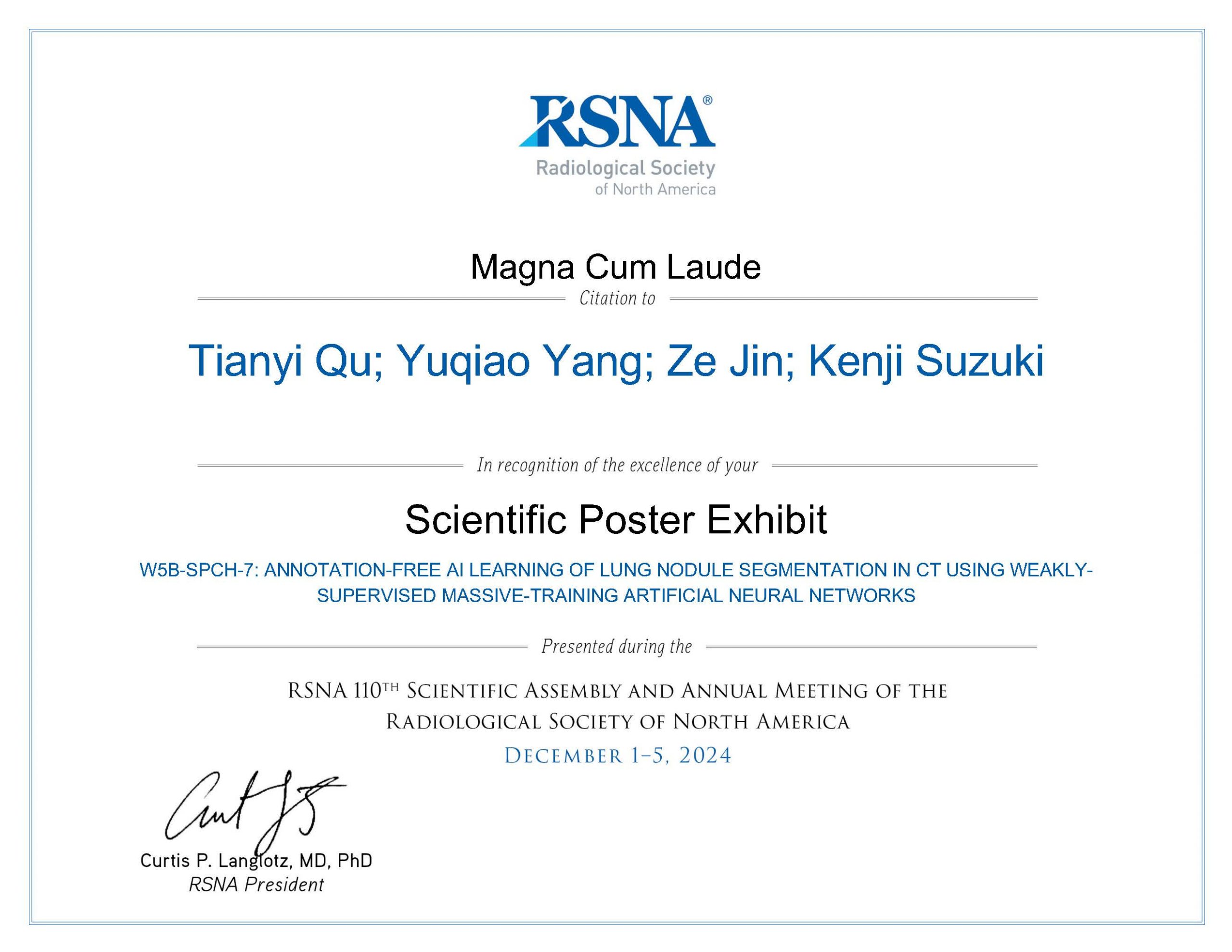
6
RSNA Science Poster Awards Cum Laude
Our study has received Science Poster Awards Cum Laude by at the 110th Scientific Assembly and Annual Meeting of Radiological Society of North America (RSNA 2024) on December 4th, 2024.
Kodera S., Chavoshian S. M., Jin Z., Watadani T., Abe O., and Suzuki K.: Super-efficient AI for lung nodule classification in CT based on small-data massive-training artificial neural network (MTANN)
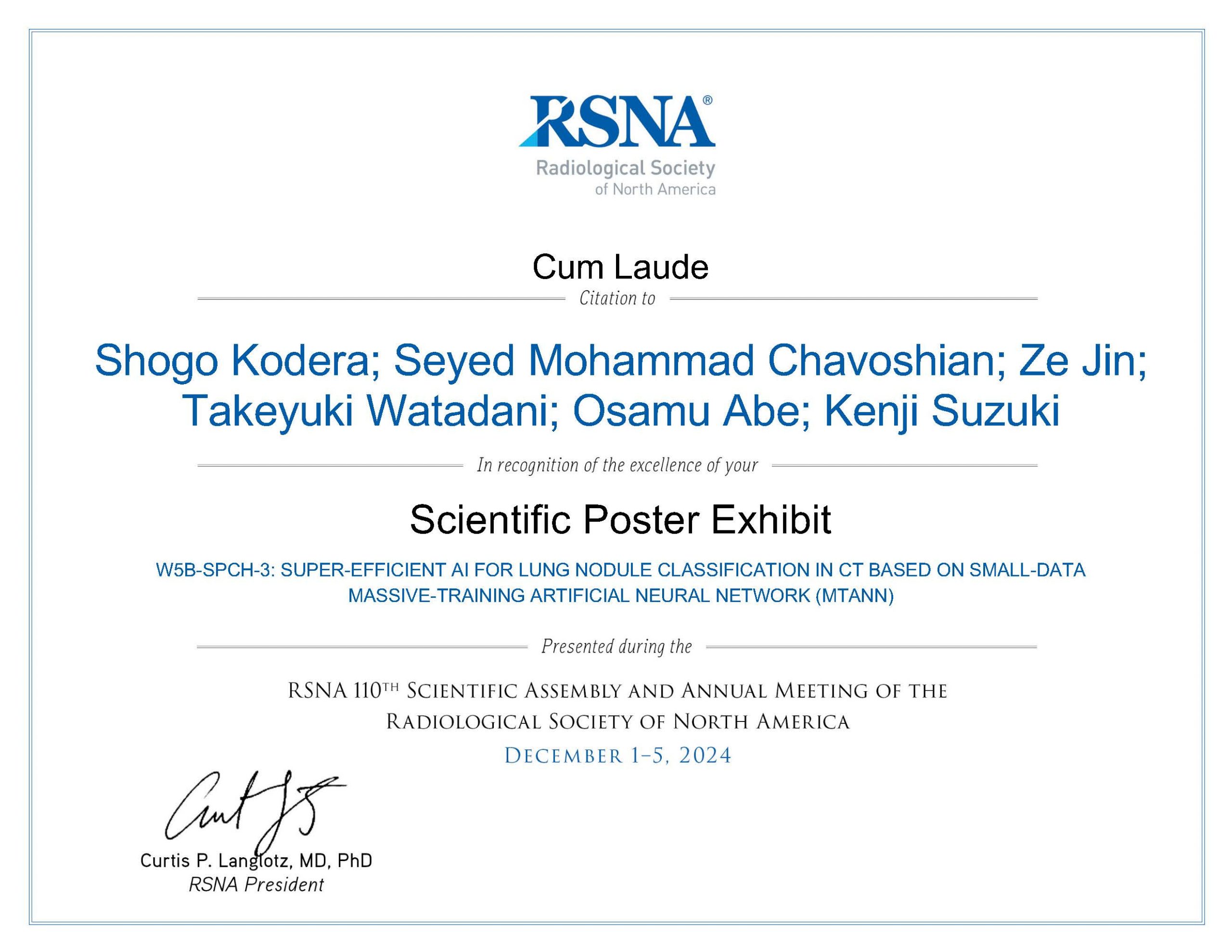
7
JAMIT Distinguished Achievement Award
Prof. Suzuki has received the Distinguished Achievement Award in recognition of his outstanding achievements in medical imaging engineering, as selected by the Board of Directors of the Japanese Society of Medical Imaging Technology (JAMIT).
Award Achievements: Pioneering research in medical image engineering using multi-layer neural networks
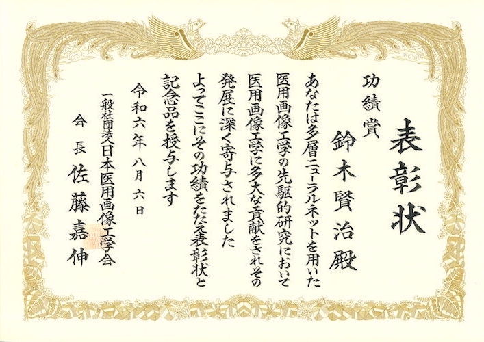
8
9
10
11
12
13
14
15
16
17
HAKUHODO Award (Best Presentation Award)
Dr. Suzuki has received the HAKUHODO Award (Best Presentation Award among 26 teams at the competition) from Hakuhodo for his presentation, “Short-term development of a variety of computer-aided diagnosis systems using small-data AI” at Startup Academia DEMO DAY 2022.
18
19
20
21
22
23
24
25
26
27